 KH Lab
Electron Micrographs of the Neuropil: Basic Literacy
Overview
Neuropil is any area of the nervous system made up mostly of unmyelinated axons, dendrites, and glial cell processes. It is dense with synapses and has relatively few cell bodies. When you look at electron micrographs (EMs) of the neuropil, you should be able to recognize dendrites, axons, and glia. You should also recognize their organelles and other structures inside them, such as the postsynaptic density (PSD), mitochondria, smooth endoplasmic reticulum (SER), spine apparatus, polyribosomes, and microtubules.
This video will help you get to know these structures: Cruise through the Hippocampal Neuropil: Brain Cells Visualized. This tutorial gives more information about synapses: Structure of Chemical Synapses.
This page is adapted from a handout by Patrick Parker, Kristen Harris Lab (2020).
Each Figure box below links a figure from the sources under References; open them while you read. The EM images on this page come from the class series.
Dendrites
The main features of a dendrite are:
- Spines that stick out from the dendrite and form synapses with axons. (Some dendrites have no spines; their synapses are on the dendritic shaft.)
- Organelles such as mitochondria and SER.
Figure: spiny dendrite, with a thin spine (S) and its synapse
Atlas Fig. 1.4.1.02. D is the dendritic shaft. You can spot the synapse by the dark postsynaptic density (PSD) in the spine head and the vesicles next to it in the axon. Scale = 250 nm. (Rat, hippocampus.) Spacek, Atlas of Ultrastructural Neurocytology. Open the figure.
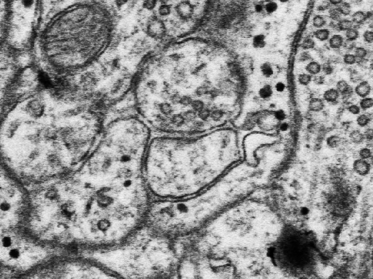
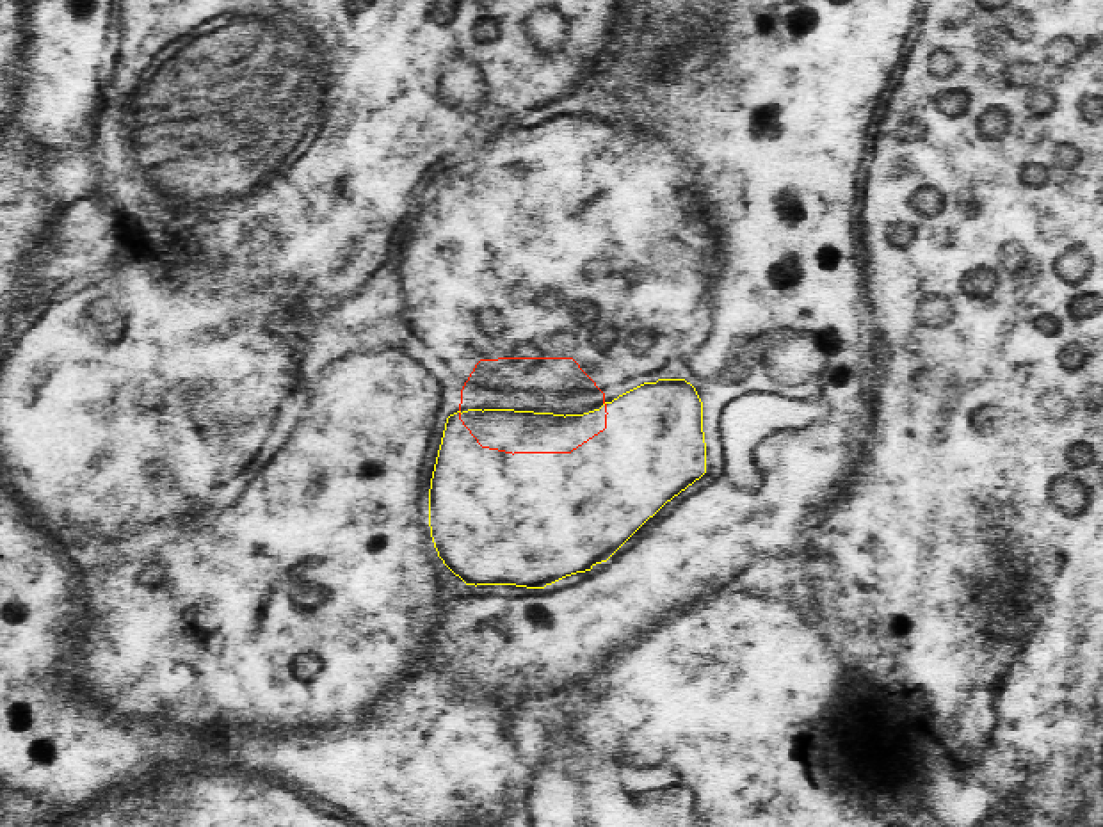
Figure: mushroom-shaped spine (s) with a spine apparatus (arrow) in its head
Atlas Fig. 1.4.1.03. D is the dendritic shaft. Scale = 300 nm. (Rat, hippocampus.) Spacek, Atlas of Ultrastructural Neurocytology. Open the figure.
Figure: 3D reconstruction of a dendrite segment (D) with spines and synapses
Atlas Fig. 1.4.1.17. T marks thin spines with macular synapses; M marks a mushroom-shaped spine with a perforated synapse. (Rat, hippocampus.) Spacek, Atlas of Ultrastructural Neurocytology. Open the figure.
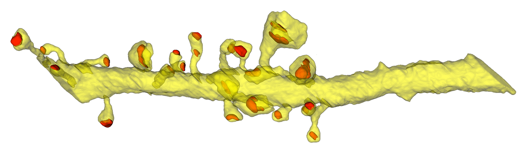
Figure: nonspiny dendrite
Atlas Fig. 1.4.06. D is the dendritic shaft, S marks shaft synapses, and the shaft holds 4 mitochondria. Spacek, Atlas of Ultrastructural Neurocytology. Open the figure.
Axons
The main features of an axon are:
- Many round vesicles in the bouton.
- Boutons that form synapses with dendrites.
- Mitochondria.
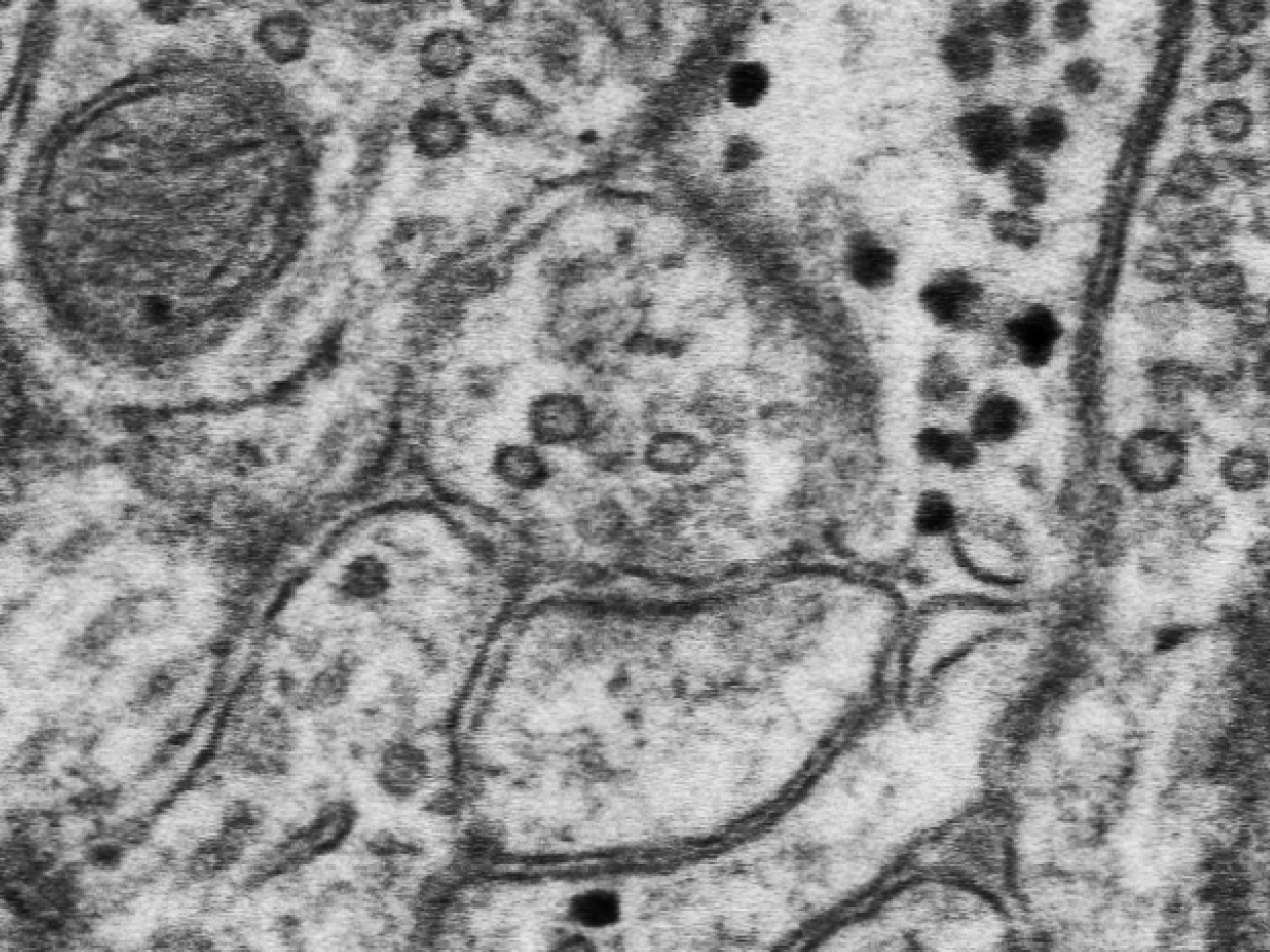
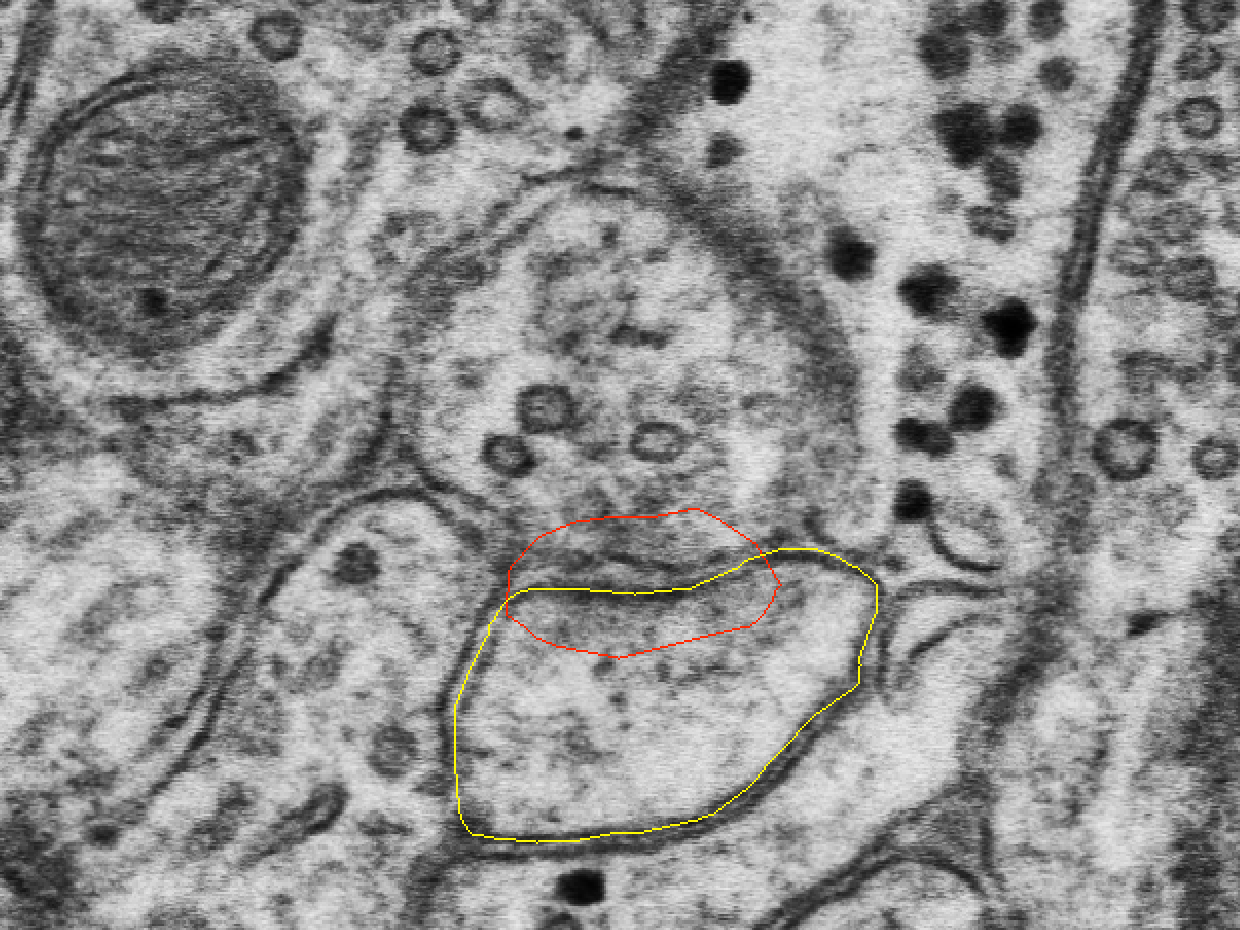
Figure: an axon swollen into an en passant bouton (B) that makes a synapse with a small spine (asterisk)
Atlas Fig. 1.5.02. Arrows mark microtubules cut along their length. Scale = 300 nm. (Rat, hippocampus.) Spacek, Atlas of Ultrastructural Neurocytology. Open the figure.
Glia
The main features of glia are:
- Pale cytoplasm.
- Dark, round glycogen granules.
- Processes that weave in among everything else: one astrocyte can fill the whole series.
Figure: astrocyte process (A) with many glycogen granules, wrapping the dendritic spines (S)
Atlas Fig. 2.1.12. The main features of the astrocyte are its pale cytoplasm, glycogen granules, and processes that weave among its neighbors. Scale = 400 nm. (Rabbit, cerebellar cortex.) Spacek, Atlas of Ultrastructural Neurocytology. Open the figure.
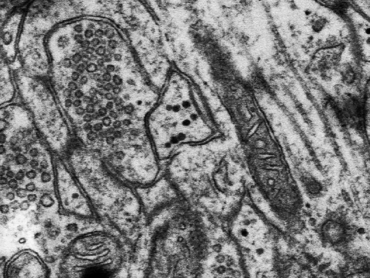
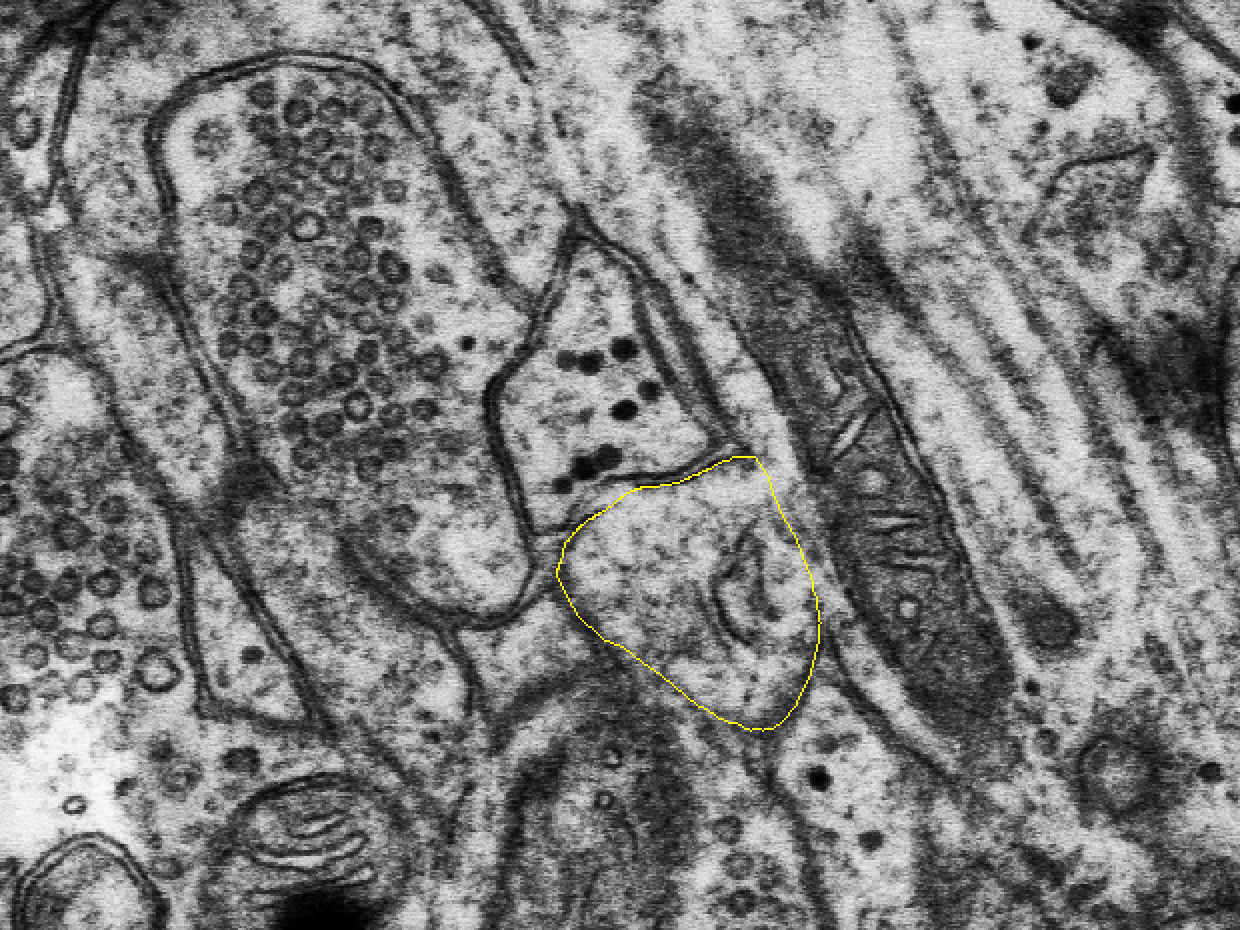
Figure: 3D reconstruction of an astrocyte
Atlas Fig. 2.1.36. Direct contacts with synaptic clefts are red. Scale = 1 µm. (Rat, hippocampus.) Spacek, Atlas of Ultrastructural Neurocytology. Open the figure.
Organelles
Mitochondria
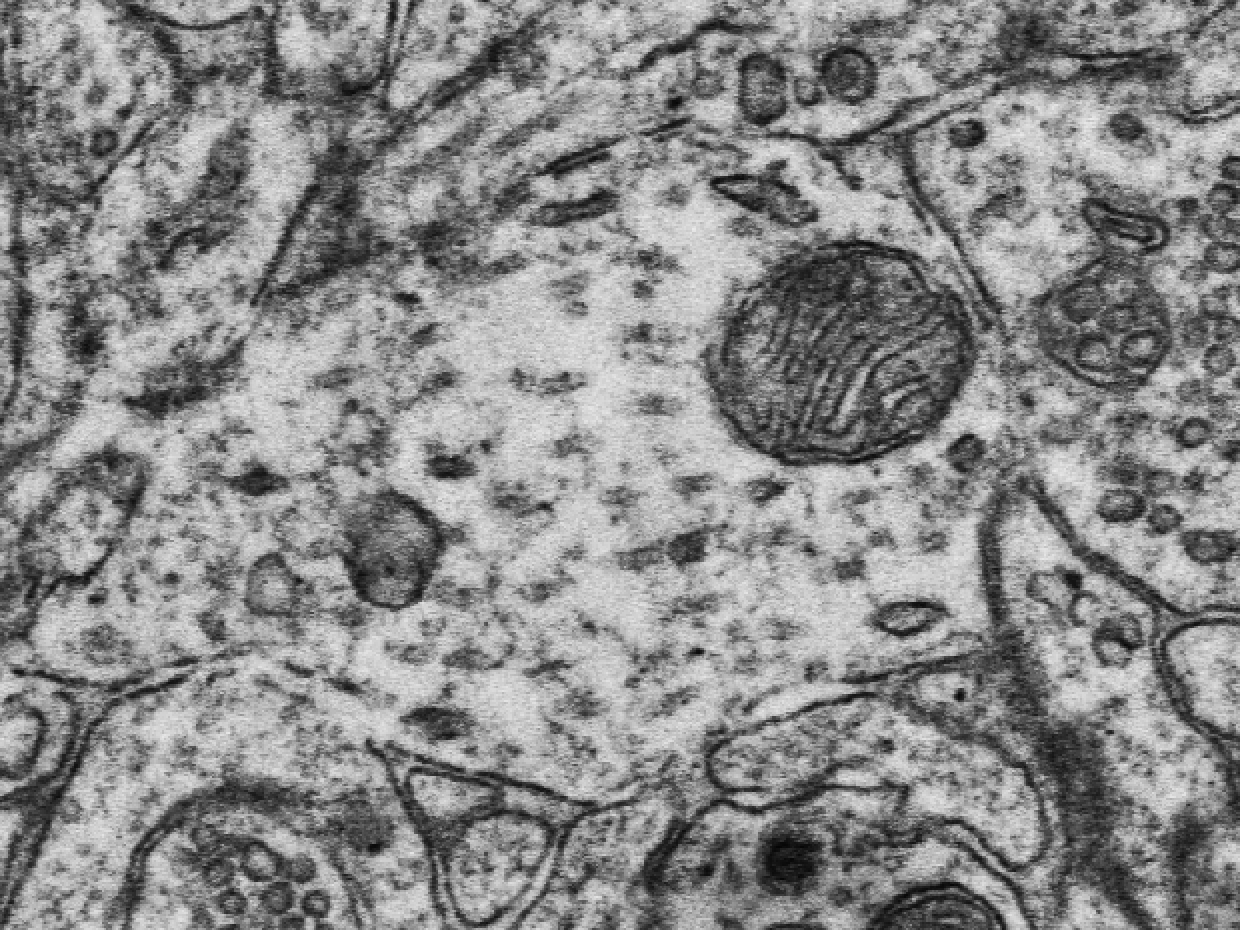
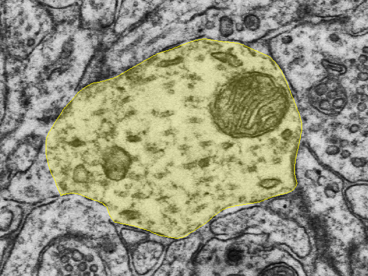
Mitochondria are large organelles that are easy to spot in dendrites and axons. A mitochondrion often runs along the length of a dendrite, and mitochondria sometimes branch.
Figure: mitochondrion in a cerebellar granule cell, cut along its length
Atlas Fig. 1.1.4.01. Spacek, Atlas of Ultrastructural Neurocytology. Open the figure.
Smooth endoplasmic reticulum (SER)
Figure: SER in a dendrite (arrowhead)
Spacek & Harris, 1997, Fig. 1. Open the paper.
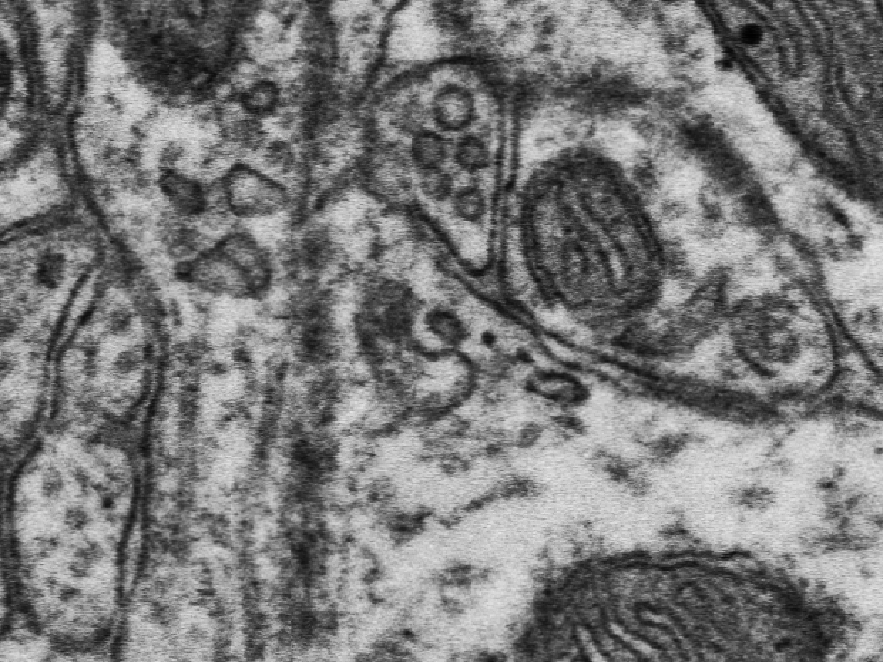
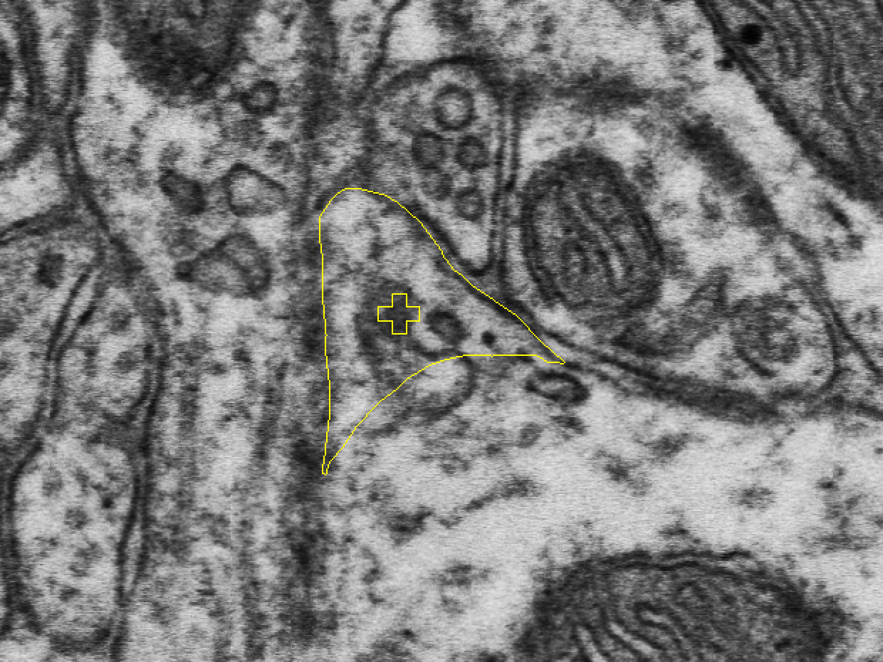
Figure: 3D reconstruction of SER in a dendrite
Atlas Fig. 1.1.2.14. (Rat, hippocampal stratum radiatum.) Spacek, Atlas of Ultrastructural Neurocytology. Open the figure.
Spine apparatus
Figure: spine apparatus cut across, with six flat cisternae and five dense plates between them
Fig. 2 of the SynapseWeb Spine Apparatus tutorial. Open the tutorial.

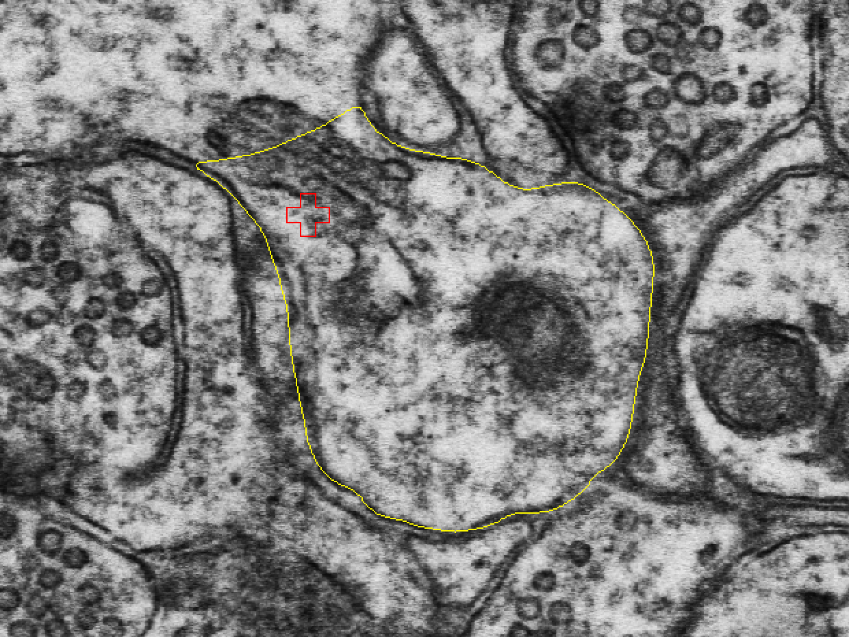
Figure: 3D reconstruction of a spine from hippocampal stratum radiatum
Fig. 6 of the SynapseWeb Spine Apparatus tutorial. A perforated excitatory synapse (red) and a macular inhibitory synapse (blue) sit on the spine head. The spine apparatus (yellow) sits in the head and neck of the spine. Open the tutorial.
References
- Spacek J. Atlas of Ultrastructural Neurocytology. SynapseWeb. synapseweb.clm.utexas.edu/atlas
- SynapseWeb anatomy tutorials. synapseweb.clm.utexas.edu/tutorials
- Harris KM, Spacek J (2016). Dendrite structure. In: Stuart G, Spruston N, Häusser M (Eds.), Dendrites, 3rd edition. Oxford University Press. doi:10.1093/acprof:oso/9780198745273.003.0001
- Spacek J, Harris KM (1997). Three-dimensional organization of smooth endoplasmic reticulum in hippocampal CA1 dendrites and dendritic spines of the immature and mature rat. Journal of Neuroscience 17:190-203. doi:10.1523/JNEUROSCI.17-01-00190.1997