 KH Lab
Lab 3.1: Smooth Endoplasmic Reticulum, Spine Apparatus, and Endosomes
Overview
In this lab you look for smooth endoplasmic reticulum (SER), spine apparatus (SA), and endosomes in 5 more spines of dendrite d03, and you place a stamp on each one you find.
- Spines: d03sp12, d03sp14, d03sp37_branched, d03sp40 and d03sp42 (see 5 additional spines).
- What you make: one stamp for each SER, SA or endosome you find, named after its spine.
- What you hand in: your saved series, uploaded to Canvas.
Each Figure box names a figure in one of the two papers under References. Open both papers before you start. The EM screenshots on this page show examples from other spines of the series, not from your 5 spines.
Smooth endoplasmic reticulum (SER)
- SER has a flattened, oval appearance surrounded by a membrane, but this shape can be variable. On one section the SER can look like a gray smudge, a small black dot, a snake-like black line, or a large vesicle-like lake.
- SER is connected across serial sections, though sometimes this continuity can be difficult to see.
Figure: snake-like tubule of SER entering a spine
Spacek & Harris, 1997, Fig. 2c. Open the paper.
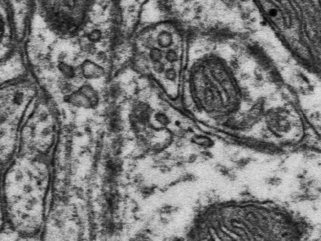
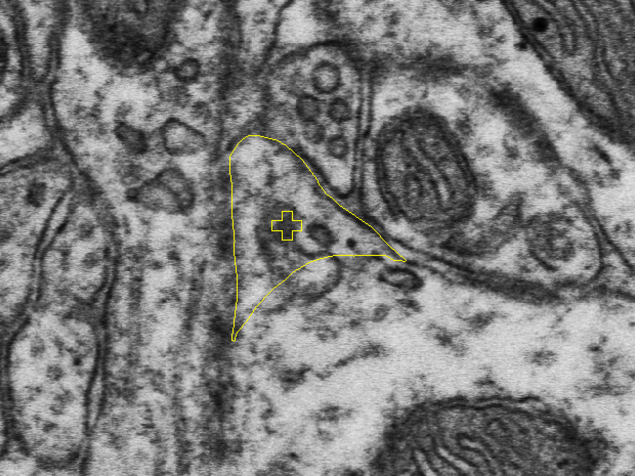
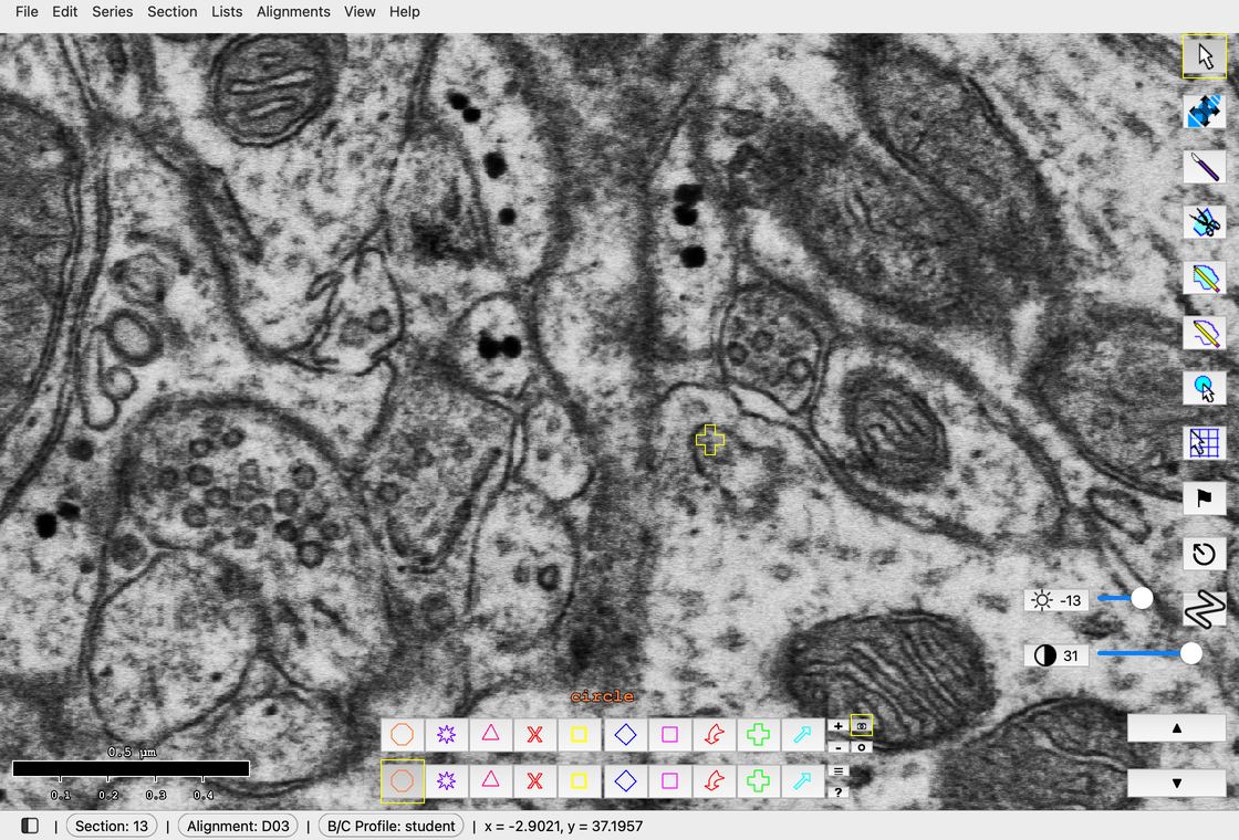
SER in spine d03sp05, sections 13 to 17, stepping up one section at a time.
The example stamps already in the series have longer names than yours: a letter after "ser" or "sa" says where the stamp sits (h for head, n for neck, s for shaft), as in d03sern05 and d03sah19. Your stamps use the short names given below, such as d03ser12.
Spine apparatus (SA)
- The spine apparatus is a specialized form of the SER.
- SA is typically seen in large mushroom spines but can also appear in the dendritic shaft.
- SA appears as flat cisternae (arrows) with electron-dense plates between them (wavy arrows); see the figure below.
Figure: spine apparatus, with cisternae (arrows) and dense plates (wavy arrows)
Spacek & Harris, 1997, Fig. 3b. Open the paper.
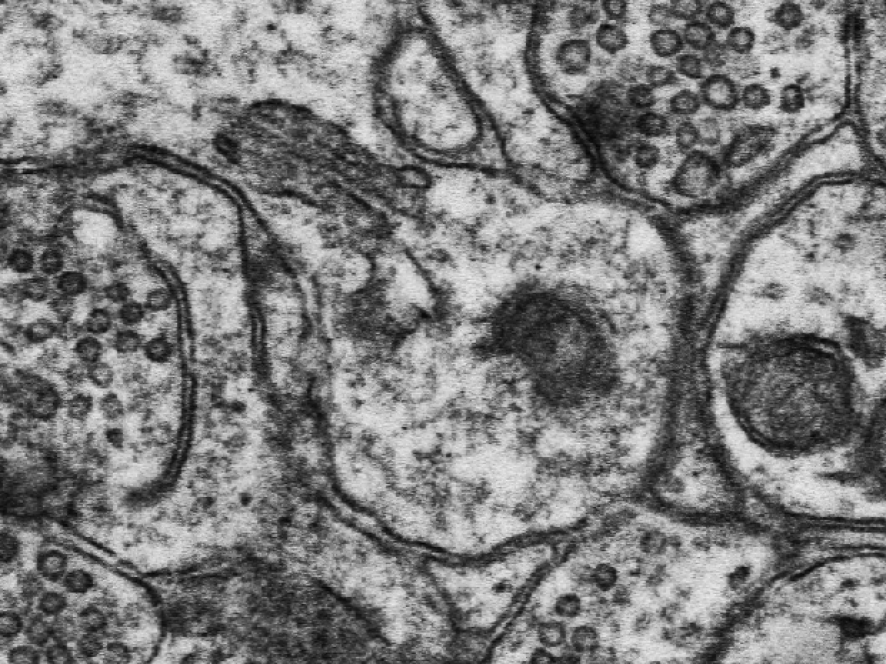
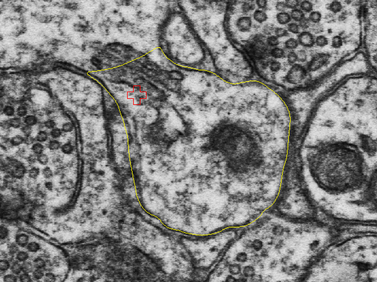
Endosomes
Endosomes are sorting organelles inside the cell. They take part in the local recycling, sorting, and breakdown of membrane and proteins.
Four major subtypes:
- Multivesicular body
- Tubule
- Smooth vesicle
- Coated vesicle or pit
Figure: drawing of endosome types in three spines
Cooney et al., 2002. Open the paper.
Endosome criteria
1. Coated vesicles and pits
- Vesicles: elliptical, with a gray interior and a clathrin coat (a coat on the cytoplasmic side made of spoke-like protrusions).
- Pits: elliptical, omega-shaped invaginations of the membrane, with gray interiors and a clathrin coat.
Figure: clathrin-coated pit, with an inset of a clathrin-coated vesicle
Cooney et al., 2002. Open the paper.
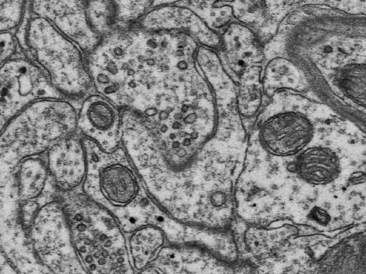
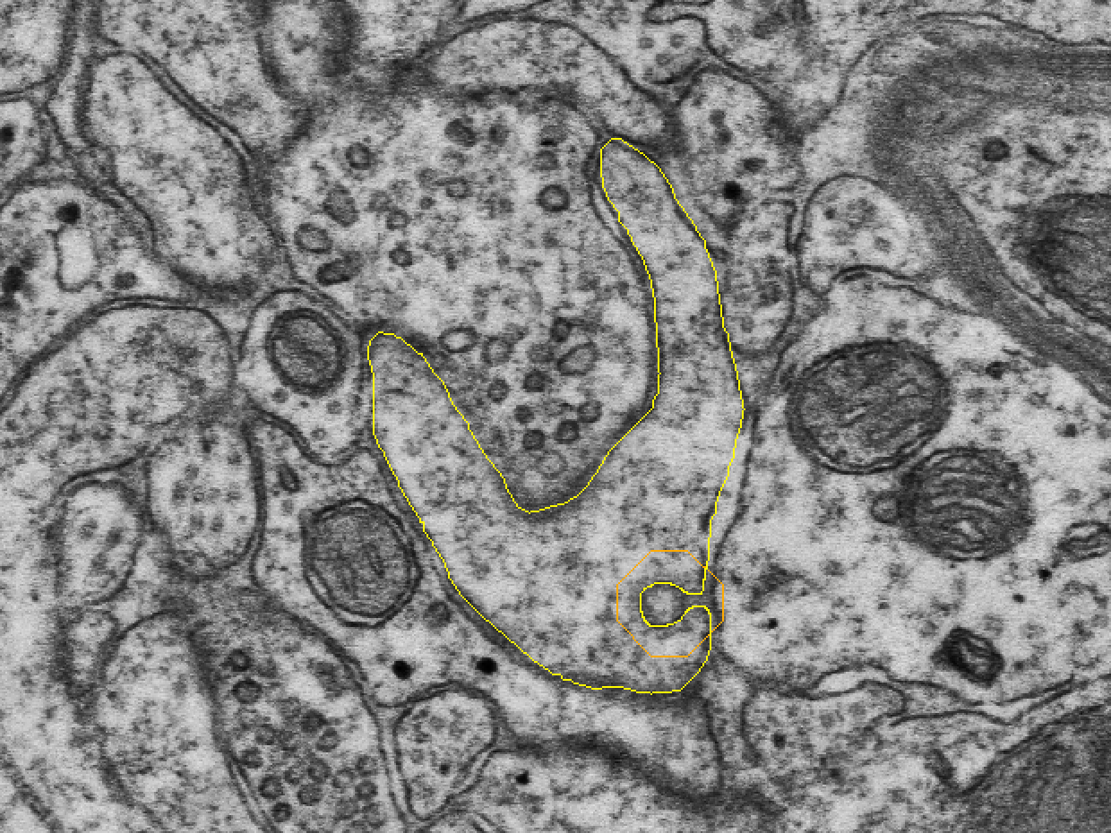
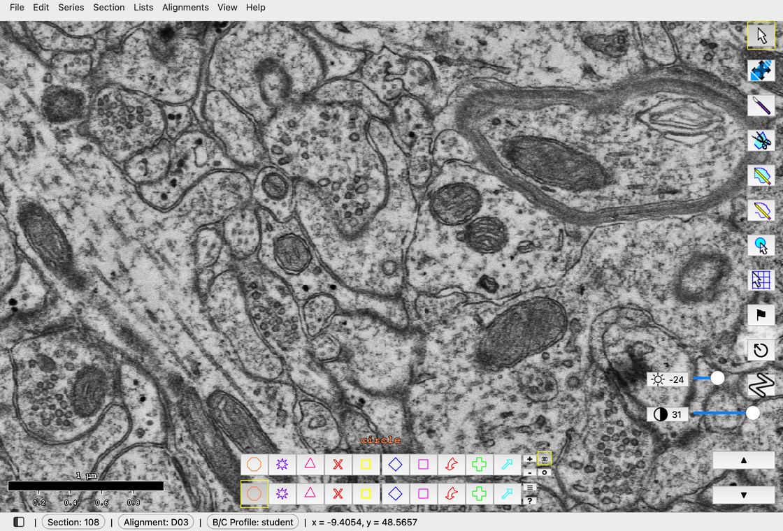
Closing in on the coated pit of spine d03sp29 on section 108.
2. Endosomal tubule
- Cylindrical compartment with a smooth outer membrane and a dark, grainy interior.
- Uniform diameter.
- May have a clathrin-like protein coat on the end, a sign that a vesicle is budding off.
Figure: endosomal tubule
Cooney et al., 2002. Open the paper.
3. Multivesicular body (MVB)
- Dark, grainy interior with a variable number of vesicles inside.
Figure: multivesicular body
Cooney et al., 2002. Open the paper.
4. Vesicles (small, large, amorphous)
- Small and large smooth vesicles: light gray interiors with a smooth outer membrane.
- Amorphous vesicles: smooth membranes, electron-lucent (pale) interiors, and irregular shapes. They often occur in clumps.
Figure: small and large smooth vesicles, and a clump of amorphous vesicles
Cooney et al., 2002. Open the paper.
5 additional spines
To learn to identify these spine resources, you are assigned 5 more spines. Each is marked by a red p-stamp, and each is already traced, with its synapse (c-trace) and contact flat area (cfa-trace):
| p-stamp (Object List group "Assigned") | Spine trace | Synapse traces |
|---|---|---|
| d03p12 | d03sp12 | d03c12, d03cfa12 |
| d03p14 | d03sp14 | d03c14, d03cfa14 |
| d03p37_branched | d03sp37_branched | d03c37a, d03cfa37a, d03c37b, d03cfa37b |
| d03p40 | d03sp40 | d03c40, d03cfa40 |
| d03p42 | d03sp42 | d03c42, d03cfa42 |
Search these 5 spines, head and neck, for SER, SA, and endosomes. To go to a spine, double-click its p-stamp in the Object List.
The group "Assigned" also holds d03p06, d03p32 and d03p47_branched, the spines you traced in Lab 1.1. They are not part of this lab.
Stamp spine SER and SA
Identify and stamp the SER or SA in each of the 5 spines. Stamp any SER and SA you see in the spine head and the spine neck.
- In the Tools window, select the Stamp tool.
- Right-click an unused palette square. The Set Attributes dialog opens.
- In Name, type the name for what you found, with ## replaced by the spine number:
- SA:
d03sa##. SA in d03sp12 isd03sa12. - SER:
d03ser##. SER in d03sp12 isd03ser12.
- SA:
- In Stamp radius (microns), type
0.05. - Click OK.
- Click on the SER or SA to place the stamp.
Use a separate palette square for each name.
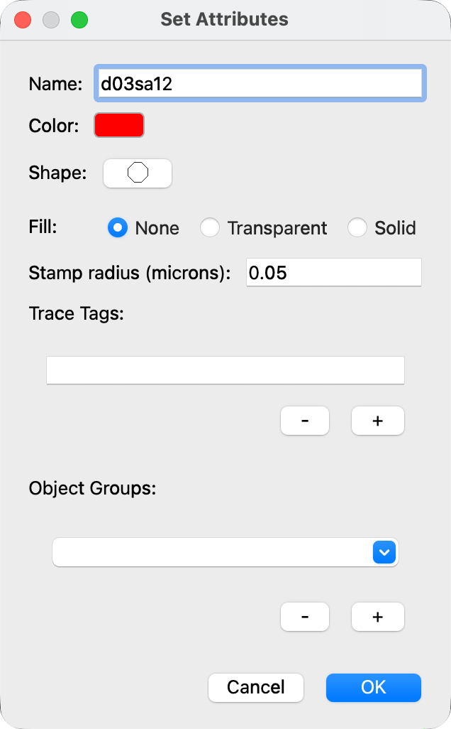
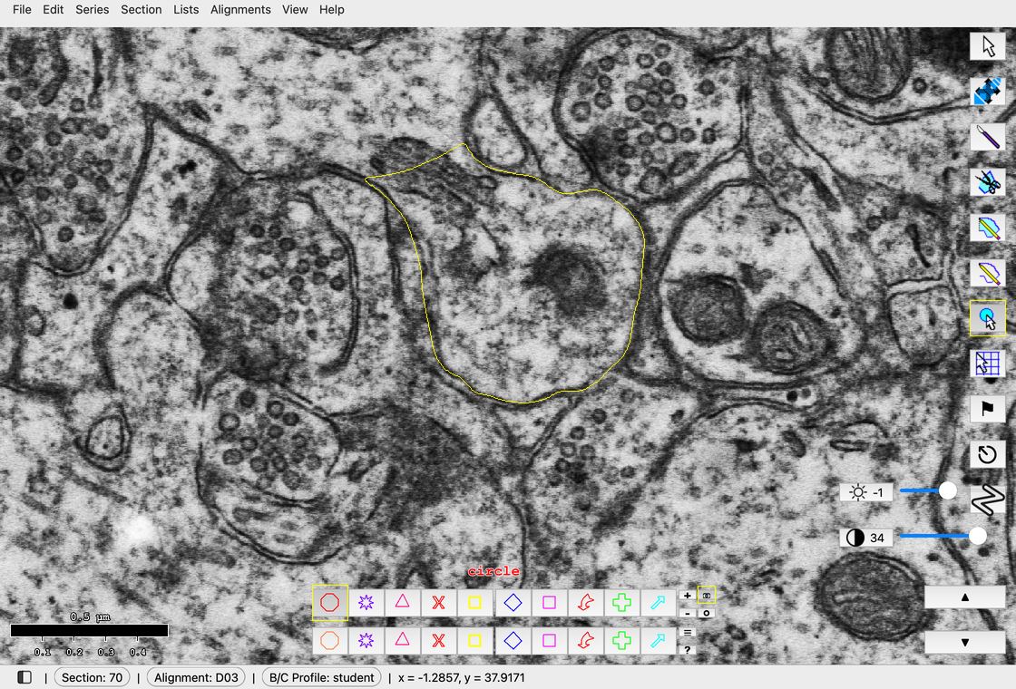
Set the name and the radius, click OK, then click on the SA. Shown in spine d03sp19, not one of your 5 spines.
Stamp spine endosomes
Look for endosomes in each of the 5 spines, and stamp each endosome you find.
Hint
You are likely to find coated pits in your assigned spines.
- With the Stamp tool selected, right-click an unused palette square. The Set Attributes dialog opens.
- In Name, type
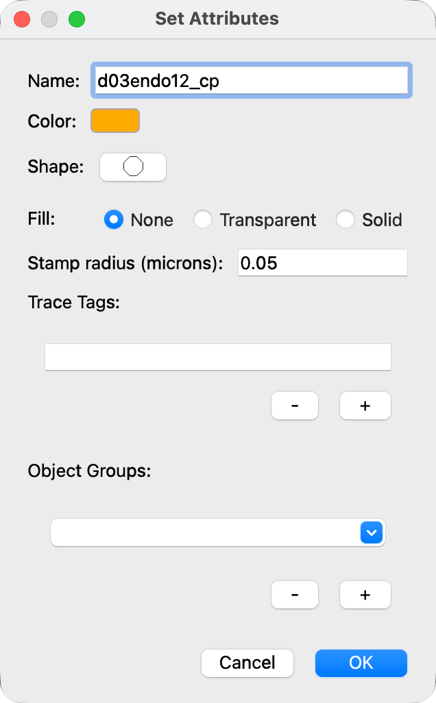
d03endo##_cp, with ## replaced by the spine number (cp stands for coated pit). A coated pit in d03sp12 isd03endo12_cp. - In Stamp radius (microns), type
0.05. - Click OK.
- Click on the endosome to place the stamp.
Check your stamps
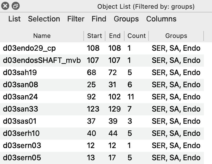
In the Object List, choose Filter > Group filter... and pick the group "SER, SA, Endo" to see the example stamps. Your new stamps are not in this group; to see them too, type a regex filter such as d03(ser|sa|endo).* in a second Object List (see Lab 1.1).
Saving
Save with Ctrl+S (Mac: Cmd+S), back up your .jser file, and upload it to Canvas, as in Lab 1.1.
References
- Spacek J, Harris KM (1997). Three-dimensional organization of smooth endoplasmic reticulum in hippocampal CA1 dendrites and dendritic spines of the immature and mature rat. Journal of Neuroscience 17(1):190-203. doi:10.1523/JNEUROSCI.17-01-00190.1997
- Cooney JR, Hurlburt JL, Selig DK, Harris KM, Fiala JC (2002). Endosomal compartments serve multiple hippocampal dendritic spines from a widespread rather than a local store of recycling membrane. Journal of Neuroscience 22(6):2215-2224. doi:10.1523/JNEUROSCI.22-06-02215.2002