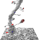
 KH Lab
Lab 2.1: Presynaptic Boutons and Mitochondria
Overview
Axons swell into vesicle-filled regions called boutons (French for "button"). This lab spans two weeks:
- Part A, Week 3: boutons and vesicles. Trace two presynaptic boutons, sort boutons into single-synaptic and multi-synaptic, and stamp the synaptic vesicles in your two boutons.
- Part B, Week 4: mitochondria. Look for mitochondria in the same boutons, and trace the ones you find.
What you work on:
- Boutons you trace: d03ax14 and d03ax32, the axons that make the synapses d03c14 and d03c32.
- Boutons you only classify and check: 8 more, listed under Identify SSB or MSB for 8 more boutons.
- What you hand in: your saved series, uploaded to Canvas at the end of each class, and your filled-in data spreadsheet.
Download the data spreadsheet
Download a copy of the spreadsheet for your bouton data and fill in the highlighted blanks:
You fill in the bouton data in Part A and add the mitochondria data in Part B. You use the spreadsheet for your Module 2 lab report.
Part A: Boutons and vesicles
Trace the presynaptic axonal boutons
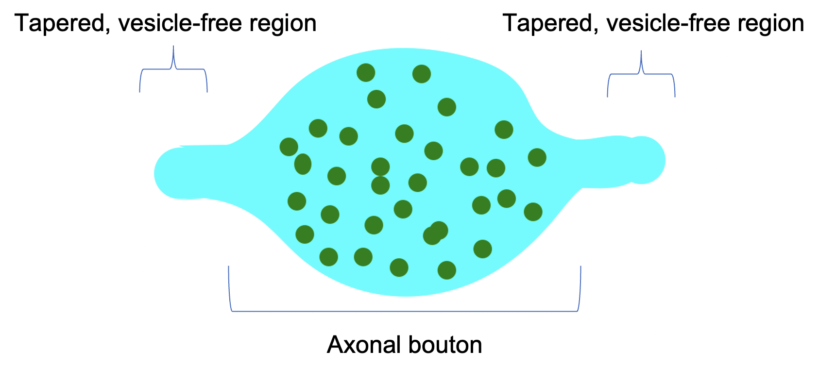
Trace the presynaptic boutons that form synapses with spines d03sp14 and d03sp32. The spines and synapses are already traced: d03sp14, d03c14 and d03cfa14 come with the series, and d03sp32, d03c32 and d03cfa32 are the traces you made in Lab 1.1 and Lab 1.2. Add the presynaptic axon, tracing only the bouton (see the cartoon below).
| Synapse (c-trace) | Contact flat area (cfa-trace) | Bouton trace | Bouton color |
|---|---|---|---|
| d03c14 | d03cfa14 | d03ax14 | cyan |
| d03c32 | d03cfa32 | d03ax32 | cyan |
Name each bouton trace d03ax##, where ## is the number of its synapse. Trace the membrane boundary of the axon, as you did for the spines.
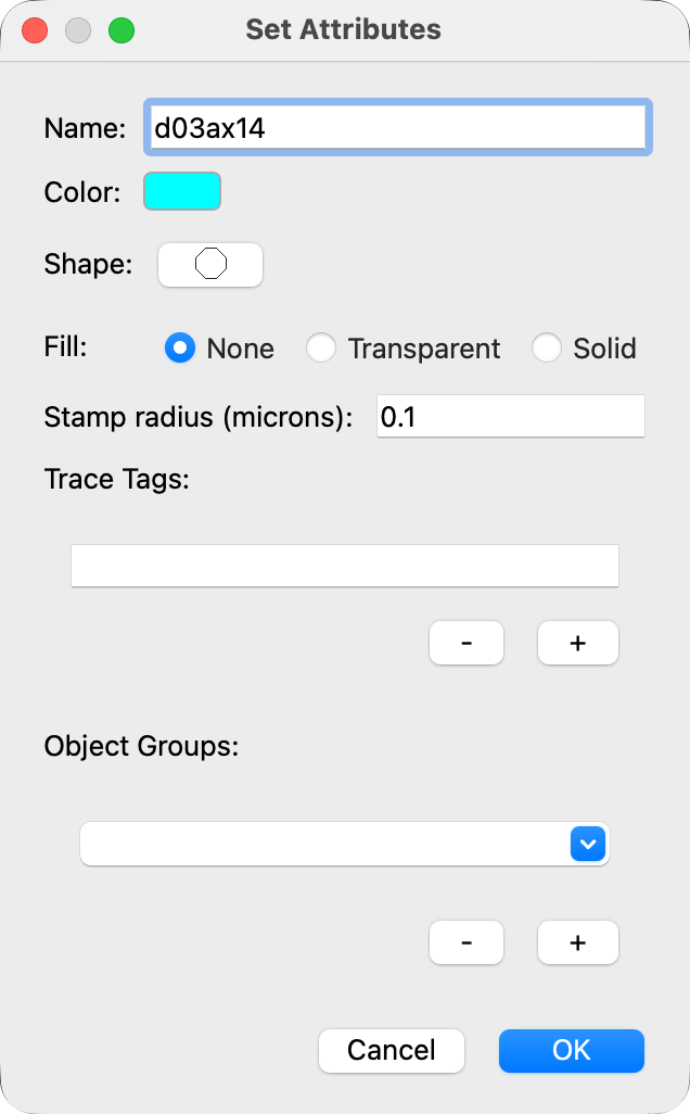
- Right-click an unused palette square. The Set Attributes dialog opens.
- In Name, type
d03ax14. - Click Color and choose cyan.
- Click OK.
- Set up a second palette square the same way for
d03ax32. - In the Tools window, select the Closed Trace tool.
- In the Object List, double-click d03c14 to go to its first section.
- Select the d03ax14 palette square and trace the outline of the axon across from the synapse.
- Go to the next section (Page Up) and trace again. Repeat on every section of the bouton.
Trace the whole bouton, up to where it starts to taper into the thinner, vesicle-free stretch between boutons. Don't trace too far! Ask your TAs if you are not sure where the bouton ends.
While you trace, watch for other synapses on the same axon.
Classify the boutons: single-synaptic or multi-synaptic
When both boutons, d03ax14 and d03ax32, are fully traced, decide for each one whether it is:
- a single-synaptic bouton (SSB), with one synapse, or
- a multi-synaptic bouton (MSB), with more than one synapse.
Look along the edges of the bouton trace for other spine heads with a PSD facing the bouton.
Hint
One of your two boutons is an SSB and one is an MSB.
Figure: an SSB and an MSB, in EM and in 3D
Bourne et al., 2013. Panels A (EM) and Ai (3D) show an SSB; panels B (EM) and Bi (3D) show an MSB. Open the paper.
Trace the additional synapse on the multi-synaptic bouton
When you have found the MSB, trace its additional synapse with a red c-trace named d03ax##c, where ## is the bouton number. For example, an extra synapse on d03ax14 would be d03ax14c. Trace it with the Closed Trace tool, as you traced the c-traces in Lab 1.2.
Hint
The MSB has only one additional synapse. Ask your TAs if you cannot find it.
Stamp the synaptic vesicles in the boutons
In your two boutons, d03ax14 and d03ax32, stamp the docked and the non-docked vesicles. Docked vesicles sit against the presynaptic membrane, facing the synapse; all the others are non-docked.
| Object | Stamp names | Color | Stamp radius (microns) |
|---|---|---|---|
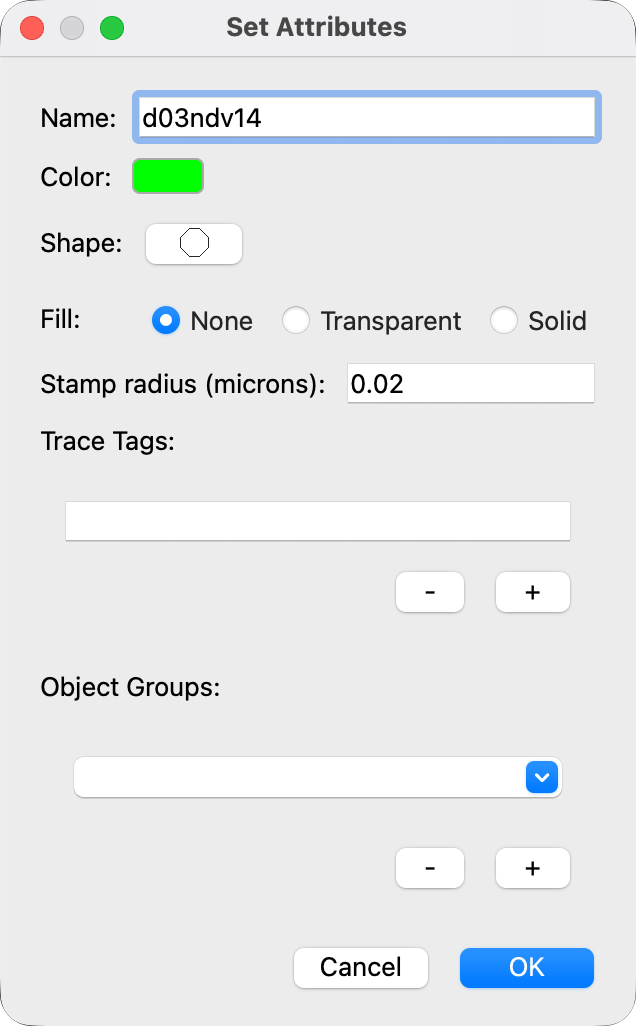
| Non-docked vesicles | d03ndv14 and d03ndv32 | green | 0.02 |
| Docked vesicles | d03dv14 and d03dv32 | magenta | 0.02 |
- In the Tools window, select the Stamp tool.
- Right-click an unused palette square. The Set Attributes dialog opens.
- In Name, type
d03ndv14. - Click Color and choose green.
- In Stamp radius (microns), type
0.02. - Click OK.
- Set up a palette square for
d03dv14the same way, in magenta. Then do the same for bouton 32. - Select the matching palette square and click on each vesicle to stamp it.
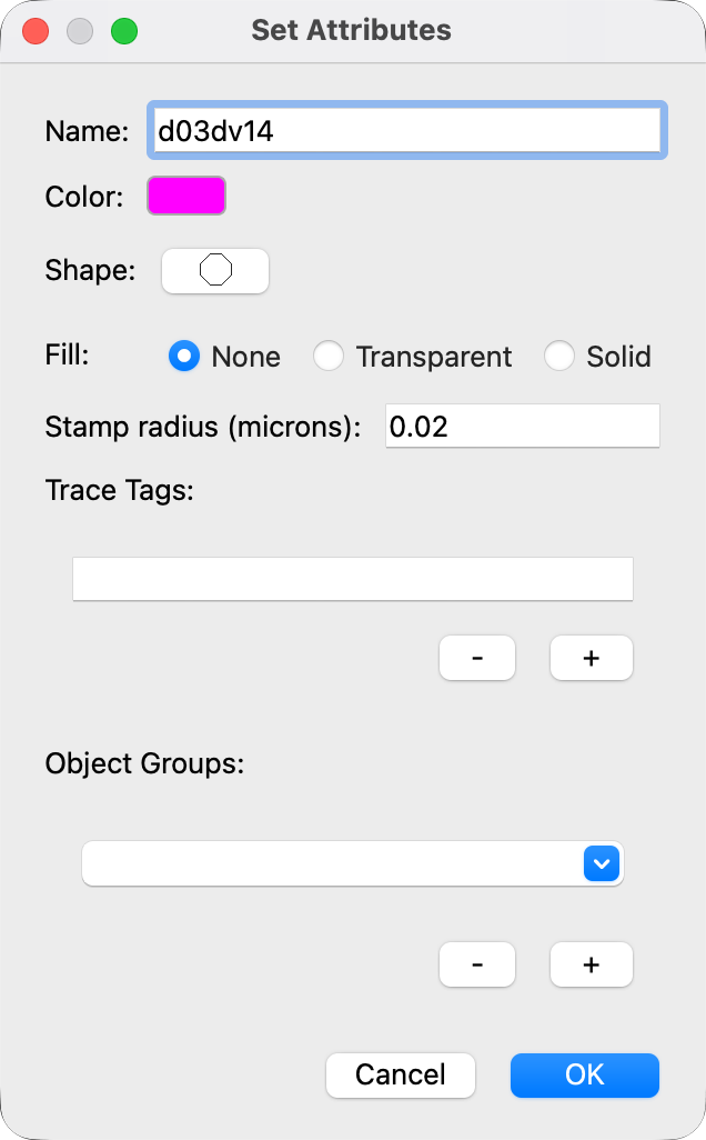
For full credit on the non-docked vesicles, your count must be within 10% of the answer key.
Hint
Think about vesicle size and shape, and about what else could be in the presynaptic bouton.
Identify SSB or MSB for 8 more boutons
For these 8 boutons you do not trace or stamp anything. Decide only whether each one is an SSB or an MSB, and record it in your spreadsheet:
- the axon at d03c06
- the axon at d03c12
- the axon at d03c15
- the axon at d03c16
- the axon at d03c40
- the axon at d03c42
- the axon at d03c47a
- the axon at d03c47b
d03c06, d03c47a and d03c47b are your own traces from Lab 1.2; the others come with the series.
These axons have no traces
These axons are not traced, so no axon name is in the Object List. Find each axon from its synapse in the Object List. The axon at d03cfa06 can still be called d03ax06, even though no d03ax06 exists. If it helps you follow a bouton through the sections, you may stamp the axon with that name.
At the end of the Week 3 class, save and upload your .jser file (see Saving).
Part B: Mitochondria
In Week 4 you look for mitochondria in the presynaptic boutons from Part A.
- Boutons you trace in: d03ax14 and d03ax32, which you traced in Part A.
- Boutons you only check: the same 8 boutons as in Part A.
Identifying mitochondria
Mitochondria are organelles that you can identify by their double membrane and the folds of their inner membrane, called cristae. The examples below show mitochondria (purple) in presynaptic boutons (cyan). The bouton and mitochondrion traces in these examples were drawn on a copy of the class series; your series does not have them.
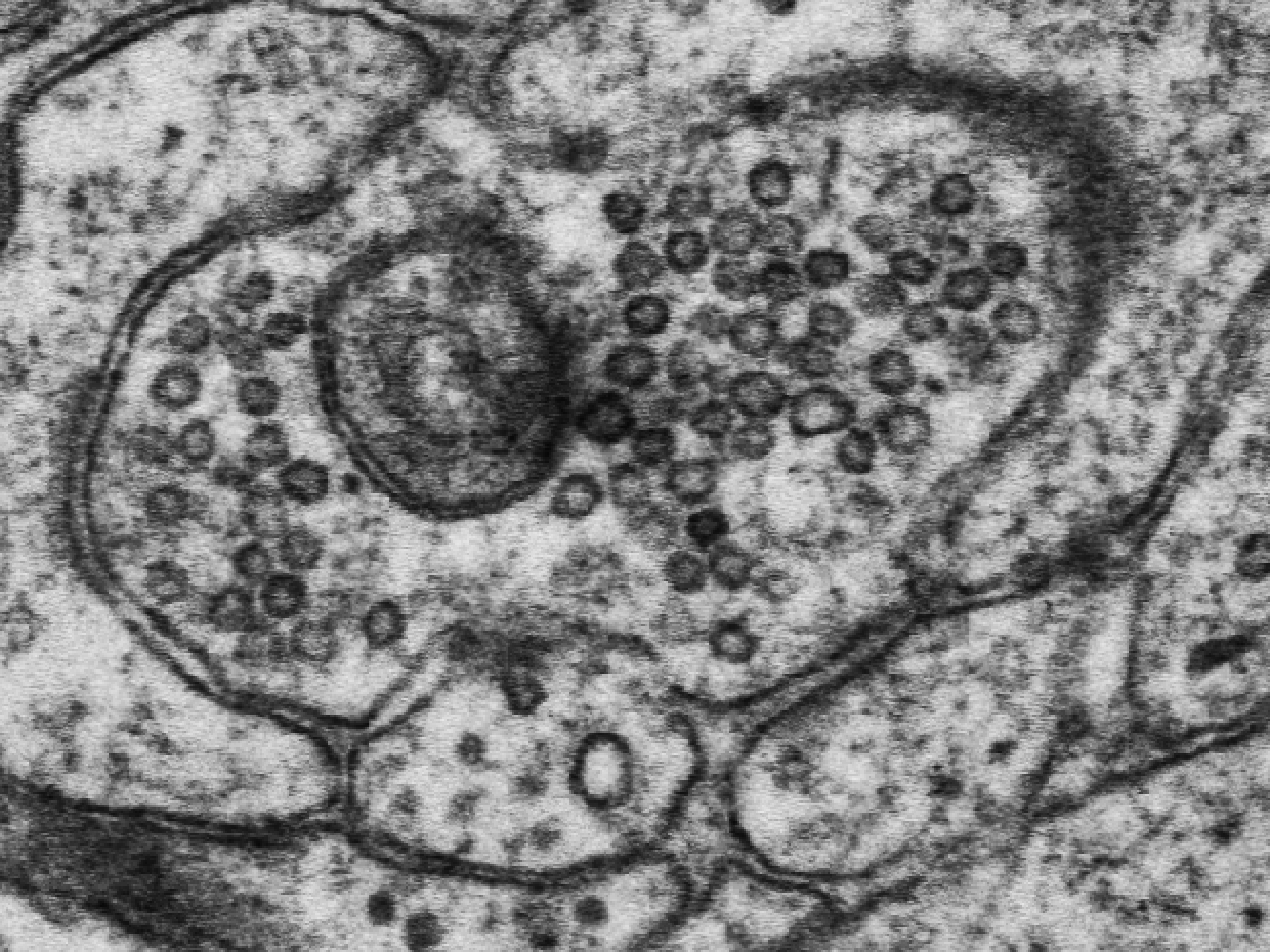
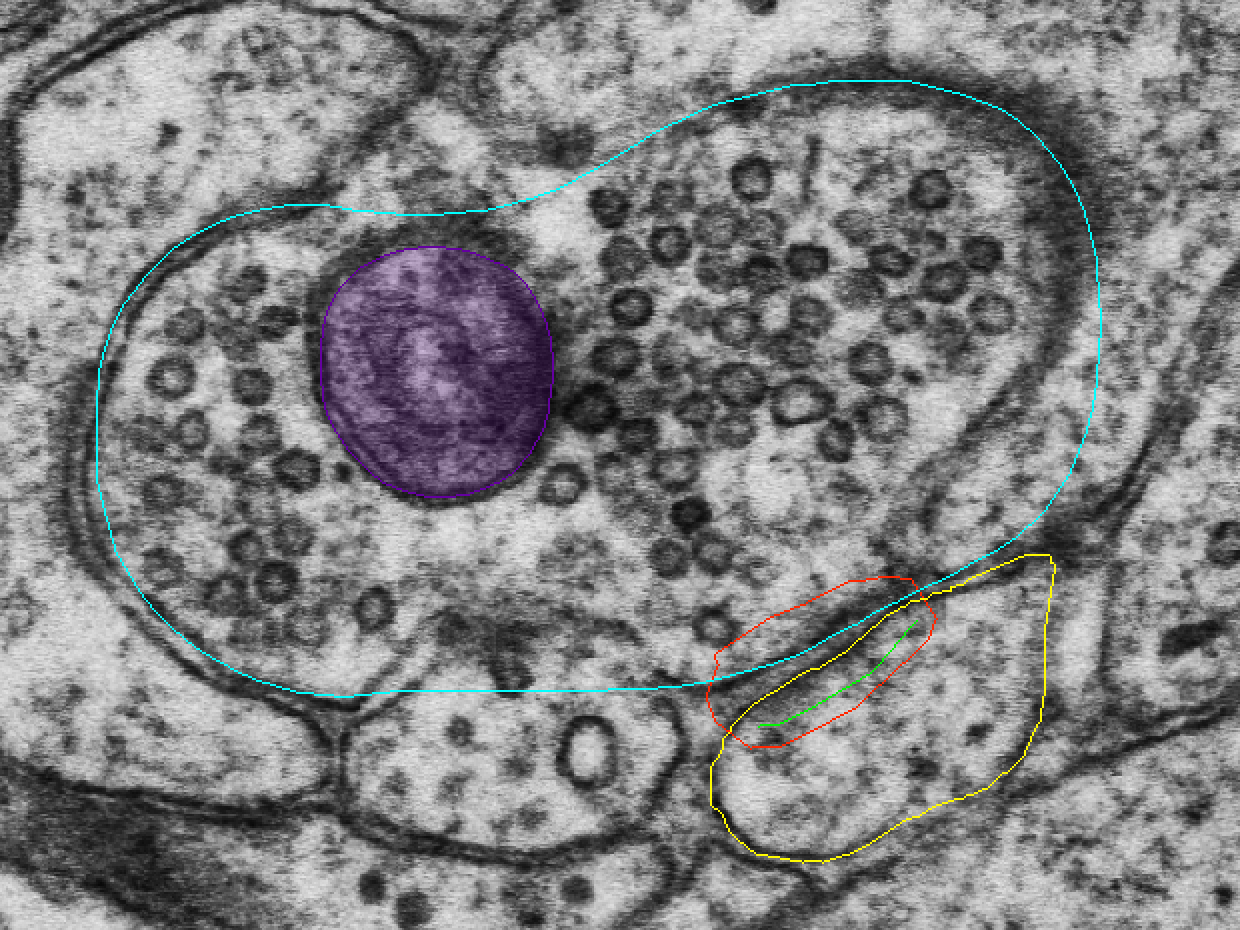
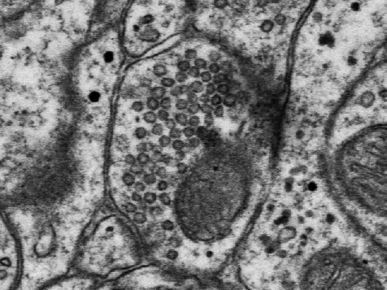
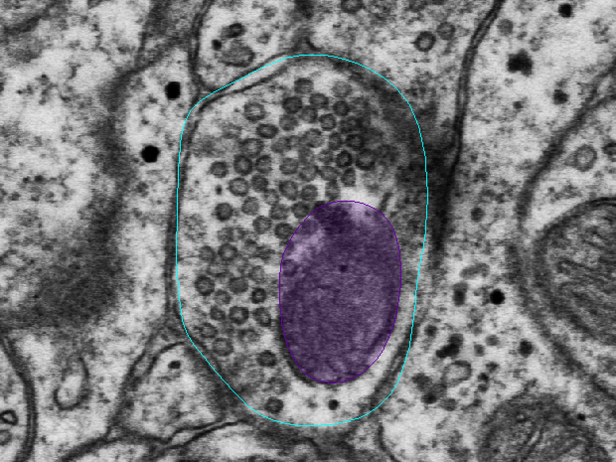
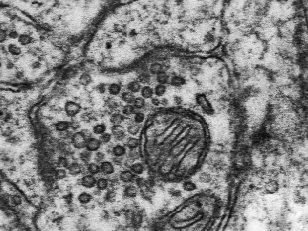
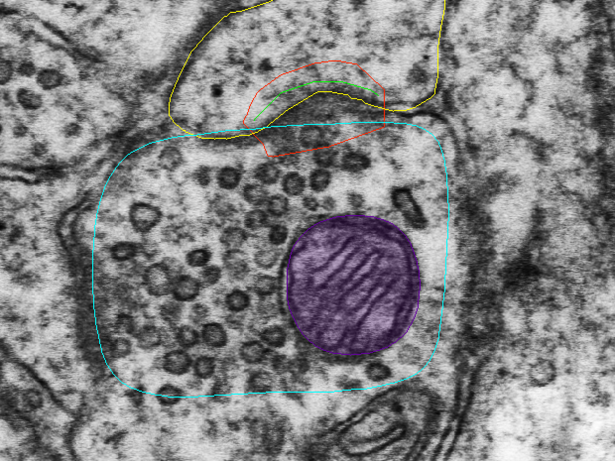
Trace presynaptic mitochondria in your two boutons
Look for mitochondria in your boutons d03ax14 and d03ax32. Trace the whole mitochondrion that lies inside the traced bouton, on every section where you see it.
Hint
Only one of the two boutons contains a mitochondrion.
- Right-click an unused palette square. The Set Attributes dialog opens.
- In Name, type
d03ax##_mito, where ## is the number of the bouton's synapse, such asd03ax14_mito. - Click Color and choose dark purple.
- Click OK.
- In the Tools window, select the Closed Trace tool.
- In the Object List, double-click the bouton (d03ax14 or d03ax32) to go to its first section.
- Trace the outline of the mitochondrion.
- Go to the next section (Page Up) and trace it again. Repeat on every section of the bouton where the mitochondrion appears.
Check for mitochondria in 8 more boutons
Your data spreadsheet has 8 more boutons without mitochondria data (highlighted in yellow). These are the same 8 boutons you classified in Part A. For these you do not trace or stamp anything. Decide only whether each bouton has a mitochondrion, and enter mito_yes or mito_no:
- the axon at d03c06
- the axon at d03c12
- the axon at d03c15
- the axon at d03c16
- the axon at d03c40
- the axon at d03c42
- the axon at d03c47a
- the axon at d03c47b
These axons have no traces
These axons are not traced, so no axon name is in the Object List. Find each axon from its synapse in the Object List. The axon at d03cfa06 can still be called d03ax06, even though no d03ax06 exists.
A synapse is hidden or will not select?
In the Object List, right-click the synapse (d03c## or d03cfa##):
- choose Operations > Unhide if it is hidden;
- choose Object attributes > Unlock if it is locked.
Update your data spreadsheet
Add the mitochondria data to your copy of the data spreadsheet: mito_yes or mito_no for each bouton, next to what you filled in during Part A. You use the whole spreadsheet in your Module 2 lab report.
Saving
At the end of each class, save with Ctrl+S (Mac: Cmd+S), back up your .jser file, and upload it to Canvas, as in Lab 1.1.
References
- Bourne JN, Chirillo MA, Harris KM (2013). Presynaptic ultrastructural plasticity along CA3→CA1 axons during long-term potentiation in mature hippocampus. Journal of Comparative Neurology 521(17):3898-3912. doi:10.1002/cne.23384