 Kristen Harris Lab
Lab W13: Perisynaptic Astroglial Processes
Overview
In this lab you work in your autosegmented D01 series. You will:
- Cut your 3 assigned spines (d01p11, d01p12, d01p13) off the dendrite at their bases and name them d01sp11, d01sp12 and d01sp13.
- Proofread the astroglia around their synapses (d01c11, d01c12, d01c13) and stamp the glycogen granules in it.
- Decide, for each of your 15 synapses, whether an astroglial process sits at the axon-spine interface.
- Fill in the spreadsheet.
The 3D views you make here can go in panels J and K of Figure 5 of your lab report: panel J is an EM of an axon-spine interface with perisynaptic astroglia and glycogen granules, and panel K is a 3D view of the spine with its perisynaptic astroglial process and glycogen granules as spheres. See Figure 5 instructions.
For any tool or menu, see the PyReconstruct Guide.
Identifying astroglial processes
You can tell astroglia by:
- Irregular shapes that weave in among the neuronal processes.
- Glycogen granules: small, dark, round dots.
- Fairly clear (pale) cytoplasm.
See also the Glia section of Electron Micrographs of the Neuropil: Basic Literacy.
Figure: astroglia at a synapse, in three examples
Witcher, Kirov & Harris, 2007, adapted. Example 1, axon-spine interface: an astroglial process (blue, with glycogen) touches both the axon (green) and the spine (yellow) next to the synapse (red PSD). Example 2, postsynaptic only: the astroglia touches the spine only. Example 3, presynaptic only: the astroglia touches the axon only. Open the paper.
Figure: 3D reconstruction of a spine (sp), its PSD, the axon (ax) with its vesicles (sv), and the astroglial process (astro) around them
Witcher, Kirov & Harris, 2007, adapted. Scale cube = 500 nm. Open the paper.
Cut your 3 spines at their bases
Your 3 assigned spines are marked by the p-stamps d01p11, d01p12 and d01p13. First, cut each spine off the dendrite at its base so you can show it in 3D with its astroglia.
Do this on every section where the spine is part of the dendrite trace:
- Press Ctrl+D (Mac: Cmd+D) to deselect all traces.
- Click the Pointer tool and click the dendrite trace to select it.
- Click the Knife tool.
- Drag the knife across the base of the spine, from one side of the dendrite outline to the other. The trace is cut in two, and both pieces are selected.
- Press Ctrl+D (Mac: Cmd+D), click the Pointer tool, and click the spine piece to select only it.
- Press Ctrl+E (Mac: Cmd+E). The Set Attributes dialog opens.
- In Name, type the spine name:
d01sp11for the spine at d01p11,d01sp12for d01p12, ord01sp13for d01p13. - Set the color to yellow and click OK.
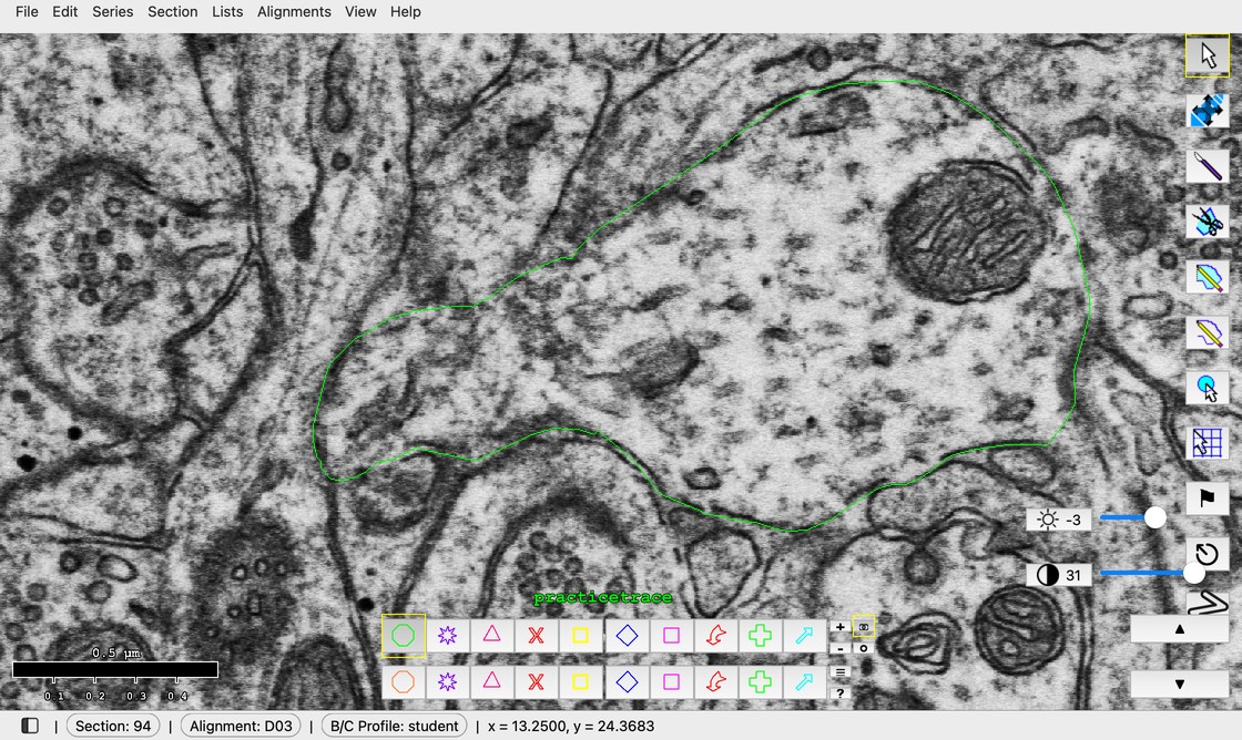
Select the trace, Knife across the spine's base, Ctrl+D (Mac: Cmd+D), select the spine piece, Ctrl+E (Mac: Cmd+E), name it and make it yellow. Shown on a practice trace around d03 and spine d03sp24 in the D03 series (section 94), with all other traces hidden.
On sections where the spine is already a separate trace, select only it and rename it the same way (steps 5 to 8).
Proofread the astroglia and stamp glycogen granules
Proofread enough of the astroglial processes around d01c11, d01c12 and d01c13 to see, in 3D, how the astroglia sits around each synapse. Fix merge and split errors with the proofreading steps from Week 10 to Week 12.
Name the astroglia
For each synapse, name the astroglia around it:
- In the Object List, right-click the astroglia object and choose Edit attributes of traces....
- In Name, type
d01g11(ord01g12,d01g13, to match the synapse). - Set the color to light blue and click OK.
Stamp the glycogen granules
- Click the Stamp tool.
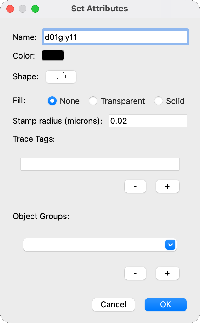
- Right-click a palette square. The Set Attributes dialog opens.
- In Name, type
d01gly11(ord01gly12,d01gly13). - Set the color to black.
- Set Stamp radius (microns) to
0.02. - Click OK.
- Click each glycogen granule inside the proofread astroglia to stamp it.
Names and 3D settings
| Synapse (red) | Spine (yellow, opacity 0.5) | Astroglia (light blue surface, opacity 0.2) | Glycogen granule stamps (black spheres, radius 0.02 µm) |
|---|---|---|---|
| d01c11 | d01sp11 | d01g11 | d01gly11 |
| d01c12 | d01sp12 | d01g12 | d01gly12 |
| d01c13 | d01sp13 | d01g13 | d01gly13 |
To set these in 3D:
- In the Object List, right-click an object and choose 3D > Edit 3D settings....
- Set 3D Type: surface for the synapse, spine and astroglia; spheres for the glycogen stamps.
- Set Opacity (0-1): 0.5 for the spine, 0.2 for the astroglia, 1 for the synapse and glycogen.
- Click OK.
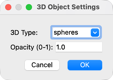
Then right-click the objects and choose 3D > Add to scene. If a new 3D type does not show, use Scene > Reload selected in the 3D window.
Is there astroglia at the axon-spine interface?
The axon-spine interface (ASI) is where the axon lies right next to the spine (Example 1 in the first figure above). If astroglia is right next to this place, where the axon meets the spine, astroglia is present at the ASI.
After you finish the 3D reconstructions of d01g11, d01g12 and d01g13, decide for all 15 of your synapses whether perisynaptic astroglia is at the ASI. Proofreading the astroglia and fixing merge and split errors will help you decide.
Follow both the spine and the axon through the serial sections, then choose one:
- YES, astroglial process at the ASI: an astroglial process sits at the ASI, next to both the axon and the spine (Example 1).
- NO, astroglial process not at the ASI: choose NO if any of these is true:
- Postsynaptic only: astroglia touches the spine only (Example 2).
- Presynaptic only: astroglia touches the axon only (Example 3).
- Presynaptic and postsynaptic, but not at the ASI: astroglia touches both the spine and the axon, but none is near the synapse.
- None: astroglia touches neither the axon nor the spine.
Fill in the spreadsheet
Fill in the yellow blanks about perisynaptic astroglia for your 15 synapses in Supplemental Table 2: Autosegmentation Outcomes, NZ Astro ASI. The spreadsheet link is on Canvas.
Save
Press Ctrl+S (Mac: Cmd+S) often, and before you close PyReconstruct.
References
- Witcher MR, Kirov SA, Harris KM (2007). Plasticity of perisynaptic astroglia during synaptogenesis in the mature rat hippocampus. Glia 55:13-23. doi:10.1002/glia.20415 (PDF on SynapseWeb)