 Kristen Harris Lab
Teaching notes, 2027 Spring
These are my notes for the teaching team on the 2027 Spring draft, which I built from the Spring 2026 Canvas course. They cover data problems to fix in the class series before it goes to students, decisions we need to make, and answers. Students never see this page.
Class project dendrites
The class project will use dendrites from the MML class dataset. The details are to come. The student pages are in Module 4 of the term index, and their open questions are under Class research project: open questions.
Examples picked for review
These are the example images where I picked the object or section myself while taking the screenshots, so you can review them in one place. All of them come from ZGBJY_crop-D03.jser. None of them shows any part of an object assigned in a Module 1-3 lab. Those are the Lab 1.1/1.2 spines and PSDs 06, 32 and 47. In Lab 2.1 they're the boutons d03ax14 and d03ax32 (synapses d03c14, d03c32) and the 8 checked boutons at d03c06, c12, c15, c16, c40, c42, c47a and c47b. In Lab 3.1/3.2 they're spines 12, 14, 37, 40 and 42. I checked each view against the series traces for those names. I drew the bouton and mitochondrion outlines by hand on a throwaway copy (names d03axEX1 to d03axEX3 and _mito). They aren't in the class series.
- Lab 2.1 Part B, example 1 (
05.png,06.png): the bouton presynaptic to d03c23 on spine d03sp23, section 78. It's a large bouton with a round mitochondrion beside its vesicle cluster. Neither d03c23 nor d03sp23 is in any lab. It replaces the Sp26 section 28 image (d03c07a), whose axon may have been the checked axon at d03c12. - Lab 2.1 Part B, example 2 (
07.png,08.png): the bouton presynaptic to d03c36b (on d03sp36_branched), section 121. A large oval mitochondrion sits next to the vesicle cluster. I didn't add vesicle stamps, since stamping vesicles is the Lab 2.1 Part A task. It replaces an Sp26 image of unknown source. - Lab 2.1 Part B, example 3 (
09.png,10.png): the bouton presynaptic to d03c22 on the small spine d03sp22, section 90. Its mitochondrion has very clear cristae. It replaces an Sp26 image of unknown source. - EM literacy, mitochondria (
03.png,03-clean.png): a mitochondrion in the d03 shaft on section 93, at about x 15.55, y 23.9 µm (the D03 alignment). I picked it by eye, since no mitochondria are traced in D03. Also in view are d03 and the unassigned d03sp24, d03sp25 and d03sp27. - EM literacy, glia (
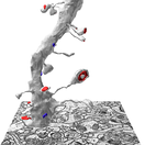
05.png,05-clean.png): a pale, angular process with a few dark round granules (about 40 nm), which I read as an astroglial process with glycogen. It's on section 100, at about x 13.85, y 23.6 µm, just above spine d03sp24 (shown in yellow). I picked it by eye, and it's the least certain pick, since no glia is traced in D03. Please confirm it's astroglia (and not, for example, a dendrite with dense granules). If it isn't, we should drop the image. - I picked these earlier, and they're already on the pages. EM literacy 01 and 04 and the Lab 1.2 PSD sections use spine d03sp28_branched with d03c28b (sections 81-85). The Top 10 merge pair uses an untraced round profile on section 163 at x 18.24, y 26.16 µm, which I picked by eye. The Guide 3D figure and EM literacy 02 use every spine and PSD except the assigned numbers.
Synapse naming in the project series
We decided on October 23 that the class uses one naming rule for synapse (c) and contact flat area (cfa) traces. Each branch gets a letter (a, b), and each synapse on one branch gets a letter plus a number (a1, a2, a3). Names only have to be unique and used the same way everywhere, and names that are easy to read are better. The rule is on Class Project: Synapse Categories.
When the MML dendrites are picked, I'll list the synapse and cfa traces that need new names in the class copies of the series (never the originals).
Source files
Everything from the Spring 2026 Canvas course is on the lab file server in /data/data1/public/Classes/NEU466G_2026_Spring/Canvas export 2026-10-22/. That includes the lecture slides, paper PDFs, the Week 10 recording, quiz and exam images, the Week 10 autosegmentation series, the Google Docs each handout came from, and every Canvas page. INVENTORY.md in that folder lists it all. I left out files with student information.
The 2027 Spring draft follows the modules, weeks, labs and grading in the Fall 2026 syllabus outline (Lyndsey Kirk, NEU 466G Fall 2026, the "Simple Syllabus" PDF). I couldn't open Lyndsey's Fall 2026 Canvas course (access denied), so I haven't used its Lab 2.1 and class project pages yet. Lab 2.1 here is the Sp26 Lab W7 and W8 merged, and the project pages are new drafts.
Getting started
- Download links. The page has the placeholder "Download link: on Canvas." The two Sp26 UT Box links on Canvas no longer work. The same files are still in my UT Box, and these links work (I checked them October 23, 2026):
- images: ZGBJY-images.zip (4.2 GB of TIFs, the Fall 2025 copy and the same file as Sp26)
- series: ZGBJY-student.jser (5.3 MB, the Fall 2025 copy and the same file as Sp26)
- current zarr dataset: ZGBJY_crop-zarr.zip (6.1 GB) and ZGBJY-studentEID.jser (4.5 MB), the Fall 2026 files. I made these two links for Spring 2027. They expire May 31, 2027. They're shared with UT accounts only, so students sign in to Box with their EID to download. You can post the links on Canvas, or we can put them on the page in place of the placeholder sentence.
- Which files students get. Sp26 handed out
ZGBJY-images(TIF folder) andZGBJY-student.jser. The current class dataset isZGBJY-crop-zarr(zarr, zip about 6.1 GB) withZGBJY_crop-D03.jser, andZGBJY_crop-D01_autoseg.jserfor W10-W13. I left the file names off the page so it works for either. Once you choose, add the names (and sizes) to step 3. - Renaming with the EID. Sp26 told students to open "ZGBJY-[studentEID].jser" and "be sure you have renamed your .jser with your EID", but no step said how or when. I left it out of the page. If you want the rename, add one step to section 3 ("Rename the .jser file to ZGBJY-yourEID.jser before you open it").
- Loaner USB drives. Sp26 said "We have loaner USB flash drives at the front of class". The page says "If you cannot download the images, ask the teaching team." Bring drives if you still offer them.
- Install. The Sp26 Miniconda +
pip install git+https://github.com/synapseweb/pyreconstruct@neu466groute no longer works, because theneu466gbranch is gone from SynapseWeb/PyReconstruct (I checked 2026-10-22). The page links to the one-click installers on pyreconstruct.org (stable v1.25.0 today) and the User Guide. Students who installed the conda version in a past term should install the app. Windows builds are unsigned, so SmartScreen may warn. The User Guide covers that. - macOS 3D scene workaround. I dropped it. The Retina/high-DPI pick bug it worked around is fixed in v1.21.1 and later, including v1.25.0. Its keyboard steps for arranging a 3D figure moved to the Guide. Please remove the "3D Scene Bug Workaround (macOS)" link from Canvas.
- I didn't link the Sp26 YouTube install tutorial (https://www.youtube.com/watch?v=HLy99ARVoBQ), since it teaches the conda route.
PyReconstruct Guide
- Credit line. The Sp26 doc says PyReconstruct was developed "with input from your TAs". The fall source and Patrick's copy say "with input from Dr. Kirk and her TAs". I used the Sp26 wording on the page. Which one do you want?
- Install and first open. The Sp26 guide had dropped the install and EID-username block. The page now sends students to Getting started for install, username and first open, and keeps a 3-step "open a series" for later weeks.
- Regex example
d01.*. I kept it as written. It matches objects in the D01 autosegmentation series (W10-W13). In the D03 series every name starts with d03, sod01.*lists nothing there. The screenshot (11.png) also showsd01.*. - Auto-save paragraph. I corrected it. The Sp26 text said PyReconstruct "does auto-save, but only to the hidden folder". It doesn't save on a timer. It keeps unsaved work in a hidden working folder and offers recovery after a crash, and the page now says that.
- New section "Arrange a 3D figure from the keyboard". I took it from the Sp26 "3D Scene Bug Workaround (macOS)" doc. The bug itself is fixed (see Getting started above).
Top 10 Things to Learn in PyReconstruct
- Cat photo. The Sp26 doc ends with "Fig. 1: UT campus cat Mashed Potato aka Smokey", a 2048 x 1977 photo with no photographer or license. I left it off the page. Add it back only with a source we're allowed to use.
- Merge images. I replaced the old stand-ins (
img/top-10-pyreconstruct/01.png,02.png) with new captures from the class series: an untraced round profile on section 163 (see Examples picked for review). - Username. "Use your UT EID as your username" is now File > Change username..., since PyReconstruct no longer asks at launch.
top-10-pyreconstruct-oldis a duplicate of the current doc (same text, same images), so there's nothing to keep.
Electron Micrographs of the Neuropil: Basic Literacy
- Source figures. I didn't embed any of the 12 figures from the handout. They come from the SynapseWeb Atlas (Spacek), Spacek & Harris 1997, or the SynapseWeb Spine Apparatus tutorial. Each one is a Figure box with a link.
- Neuron line drawing. The PDF on Canvas (
files/88030912-EM_Literacy.pdf) credits it to www.newworldencyclopedia.org/entry/Dendrite and gives no license, so I left it off the page. - Class-dataset images. SER and SA use the Lab 3.1 examples (d03sp05 section 15, d03sp19 section 70). Five more are now on the page (spine with PSD, 3D dendrite, shaft mitochondrion, bouton, astroglia). I picked the mitochondrion and astroglia by eye, since no mitochondria or glia are traced in D03. See Examples picked for review.
- Assigned-object check. The examples use d03sp05, d03sp19 and d03sp24, and none of them is in the D03 "Assigned" group. Please confirm they aren't assigned in any Sp27 lab (Lab 2.1 boutons and mitochondria, and the extra labs W4 caliber and W9 inhibitory) before the pages go to students.
- Author credit. The page says "adapted from a handout by Patrick Parker, Kristen Harris Lab (2020)", since both source copies name Patrick. Patrick, let me know if you'd rather I remove the credit.
Lab 1.1: Dendritic Spines
The assigned spines in this lab are d03sp06, d03sp32 and d03sp47, traced from p-stamps d03p06 (sections 18-27), d03p32 (119-122) and d03p47_branched (177-181). None of them is in the class series yet.
These need fixing in the class series (ZGBJY_crop-D03.jser) before it goes to students.
- There's no
Module_1group. The Sp26 handout says to filter by Module_1. The page says Dend+Spines (82 objects: d03, 42 non-shaft p-stamps and 39 finished spines), which matches the existing image and clip. Do you want aModule_1group with d03p06, d03p32 and d03p47_branched (then we retake the Object List image and clip and change the page), or keep Dend+Spines? I'd keep Dend+Spines, since it needs no change to the series. - The palette has no d03sp06, d03sp32 or d03sp47 squares. It's the stock 20-shape palette. The page says they "are already set up for you" and gives a fallback for a missing one. We should either add the three squares (yellow [255, 251, 0], Fill None) to the series, or change that sentence to "Set up a palette square for each spine". Optionally, we can also put Dend+Spines in each square's Object Groups field. Then new spines show up in the group-filtered Object List, and students don't need the second list.
- There's a stray closed trace named
circle(orange, no group) on section 163, the section the series opens on. It's the first row of the unfiltered Object List, and it shows in images 01 and 07b and the Lab 1.2 cfa-regex clip. We should delete it and then retake those images. - The stamp is d03p47_branched, but the handout names the spine d03sp47. Every other branched spine in the series keeps the suffix (d03sp07_branched, d03sp28_branched ...). Do we keep d03sp47 (Lab 1.2 uses d03sp47 branch a/b, d03c47a/b), or switch both labs to d03sp47_branched?
What I couldn't check:
- "One branch of d03p47_branched goes a long way." No copy of the series has d03sp47 traced.
What I changed on the page from the Sp26 doc:
- I dropped the note "these are example Groups ... from Fa25".
- The Open Trace warning no longer says students must redo their work, since the Trace List's right-click menu has Set closed (current section only).
- I added Backup now (Ctrl+Shift+B) to the attendance step and "Click Yes" to crash recovery.
Lab 1.2: The Postsynaptic Density
The assigned traces in this lab are d03c06/d03cfa06, d03c32/d03cfa32, d03c47a/d03cfa47a and d03c47b/d03cfa47b, on the Lab 1.1 spines. None of them is in the class series yet.
- Hidden PSDs and example stamps. In
ZGBJY_crop-D03.jser, every finished c and cfa trace, every polyribosome stamp and every SER, SA and endosome stamp is stored hidden (d03sah19 on one of its five sections). No spine trace is. Students see none of them until they press A or unhide them, so the Lab 1.2 PSD examples and the Lab 3.1 and 3.2 example stamps depend on it. Do we unhide them before release (Series > Hide > Unhide all traces (entire series)) or keep them hidden on purpose? I'd unhide them, since the pages show them. - The five cross-section panels (images 11-15) now come from the class series: d03sp28_branched / d03c28b / d03cfa28b on sections 81-85, and the page says so. They replace the Sp26 panels from series CLZBJ (d04p03). That spine isn't assigned in any lab.
- The Sp26 doc dropped a sentence the Fa25 doc had under Oblique: "The PSD will appear as a larger smear and you will not see the extracellular space in the synaptic cleft." I followed Sp26 and left it out. If it was dropped by accident, I'll put it back.
- The Sp26 doc changed the meaning of en face from "facing forward" or "in front" (Fa25) to "opposite" or "across the street". I used the Sp26 wording on the page.
- The Sp26 doc's "Sections 3, 4, and 5: Trace as above" contradicts its own section-5 image (no PSD). I used the Fa25 text on the page, where section 5 has no PSD.
- I added a numbered "Trace one PSD" block (Open Trace for the cfa line, then Closed Trace for the c-trace, on each section) and the Fill settings shown in the images (c: Transparent, cfa: Solid). The finished c-traces in the series use Fill None. Either works.
- The regex
d03cfa.*lists the 46 finished cfa objects plus the student's four. To list only the student's, used03cfa(06|32|47).*.
Lab 2.1, Part A: Presynaptic Boutons and Vesicles (Sp26 Lab W7)
I merged Sp26 Lab W7 and Lab W8 into one page, Lab 2.1, over Weeks 3 and 4 (Part A boutons and vesicles, Part B mitochondria), as in the Fall 2026 plan. I kept every fix, image and open question from both. The two sections here are the W7 and W8 notes. The Saving section is shared, and the data spreadsheet is linked once.
The assigned objects, which never appear in an example image, are boutons d03ax14 and d03ax32, their vesicle stamps, and the MSB's extra synapse (d03ax14c or d03ax32c). Students only classify the axons at d03c06, d03c12, d03c15, d03c16, d03c40, d03c42, d03c47a and d03c47b, and the answers are in the spreadsheet.
- Vesicle names and colors. The Sp26 doc gives three versions. In the prose they're d03ndv## (green) and d03dv## (orange). In the table they're d03ax14_ndv / d03ax32_ndv (green) and d03ax14_dv / d03ax32_dv (magenta). In the screenshots they're d03ndv14 (green) and d03dv14 (magenta). The page uses d03ndv## / d03dv## in green / magenta. Do you want to keep that? I'd keep it, since it matches the answer-key screenshots and the d03 + type + number pattern, and fix the answer key if it uses the table's names.
- Docked-vesicle definition. The doc never defines "docked". I added this to the page: "Docked vesicles sit against the presynaptic membrane, facing the synapse; all the others are non-docked." Please confirm it. Also, does a vesicle seen on two sections get one stamp or two? The 10% count rule depends on it.
- Half the synapses are the students' own work. d03sp32, d03c32, d03cfa32 (traced bouton) and d03c06, d03c47a, d03c47b (classified boutons) aren't in the class series. They're the students' Lab 1.1 and 1.2 traces. A student who missed or misnamed them needs a TA's help or a copy of the answer series. The page says which ones are theirs.
- Answers (from the doc). One of d03ax14 / d03ax32 is an SSB and the other is an MSB, and the MSB has one extra synapse. I kept both hints as in Sp26.
- Spreadsheet link. The page links the Sp26 Google Sheet
1l2j6iz9jaOzdlgdMg1k_pbpMZo_iReGbnH3RYQ6b-tU(Sp26 W8 linked it again). I haven't opened it. Before release, please check that it's the 2027 template, has no answers or student data, and is view-only (the page says "download a copy"). - Bourne et al. 2013 figure. The doc says "adapted from" without a figure number. The page cites the paper and names the panels (A, Ai, B, Bi). If you know the figure number, I'll add it.
Lab 2.1, Part B: Presynaptic Mitochondria (Sp26 Lab W8)
The assigned objects, which never appear in an example image, are d03ax14_mito / d03ax32_mito and the boutons d03ax14, d03ax32. Students only check the axons at d03c06, d03c12, d03c15, d03c16, d03c40, d03c42, d03c47a and d03c47b, and the answers are in the spreadsheet (mito_yes / mito_no).
- Example images replaced (2026-10-22). All three examples are now new captures from the class series with known sources: d03c23 (section 78), d03c36b (section 121) and d03c22 (section 90). None of them is in either list. See Examples picked for review. The rest of this item is about the old Sp26 images.
- Old Sp26 images. All six come from a teaching-team series with traced axons. Five carry PyReconstruct's hover label of the closest trace, "d03ax12" (one also has "d03ax17a"). The axon at d03c12 is one of the 8 checked boutons.
- I located example 1 at section 28, x 4.16, y 6.26 µm. Its bouton synapses with d03c07a on d03sp07_branched, which is in neither list. Please confirm that this axon isn't also the axon at d03c12 (the label suggests the answer series named it d03ax12). If it is, the image shows that axon with a mitochondrion and should go.
- I couldn't locate examples 2 and 3 in the class series (no match on any section). They may be outside the crop. By eye they don't match the d03c14 synapse. Please confirm they aren't the d03ax14 or d03ax32 bouton (answer: only one has a mitochondrion) or one of the 8 checked boutons. Example 2 also shows about 50 green vesicle stamps, which would be a Lab 2.1 Part A answer if it were d03ax14 or d03ax32.
- They're no longer on the page.
- Answer (from the doc). Only one of d03ax14 / d03ax32 contains a mitochondrion. I kept the hint on the page.
- TA tip. I changed "Right click > Object > Unlock" and "Operation > Unhide" to "Object attributes > Unlock" and "Operations > Unhide" in the Object List's right-click menu. Nothing is locked in the class series, but every PSD and example stamp is stored hidden (see Lab 1.2), so Unhide matters from Lab 1.2 on, not only after a student hides something.
- Student-made objects. Same as in Lab 2.1 Part A. d03ax14 and d03ax32 come from Lab 2.1 Part A, and d03c06, d03c32 and d03c47a/b come from Lab 1.2.
- Spreadsheet link. It's the same Google Sheet as Part A, and the merged page links it once (the
?usp=sharinglink). The Sp26 sheet is titled for Figure 3, an Sp26 assignment. The page calls it the "Bouton data spreadsheet" and says it's for the Module 2 lab report. Please rename the 2027 copy to match.
Lab 3.1: Smooth Endoplasmic Reticulum, Spine Apparatus, and Endosomes
The assigned objects, which never appear in an example image, are spines d03sp12, d03sp14, d03sp37_branched, d03sp40, d03sp42 and every SER, SA and endosome stamp in them.
- Stray example stamp. The 2026-fall staff note still applies to the current
class series (
ZGBJY_crop-D03.jser, modified 2026-10-18). The SER stampd03sern44(group "SER, SA, Endo", sections 165-169) sits inside spined03sp42on all five sections. d03sp42 is one of the five assigned spines, so students see one answer before they start, and the name points to spine 44, not 42. We need to delete the stamp or move it to a spine that isn't assigned before the series goes to students. No other example stamp (SER, SA, Endo or Polyribosomes groups) lies inside the five spines. - Hints and extra credit dropped in Sp26. The Sp26 doc no longer says "Each of the 5 assigned spines will have either SER or SA", "All but one of these spines has at least one endosome", or "There may be additional endosomal structures that need a second stamp", and it dropped the +0.25 extra credit for finding every coated pit. I followed Sp26 on the page. If 2027 keeps any of them, I'll put them back.
- The branched spine's names. For d03p37_branched, "##" could be 37, 37_branched, or 37a/37b (its synapses are d03c37a and d03c37b). The page says only "## replaced by the spine number". Which do you want? I'd tell students d03ser37a / d03ser37b, to match the synapses.
- Endosomes other than coated pits. The page only gives the
_cpsuffix. If a student finds an MVB, tubule or vesicle, there's no name for it. The series has one example,d03endosSHAFT_mvb. Do we pick a suffix (_mvb,_tub,_ves?) or say "stamp coated pits only"? - Example stamp sizes. The example stamps aren't 0.05 µm, and students may ask why. In radius, d03sah19 is 0.044, d03sern05 0.032, d03sern44 0.020 and d03endo29_cp 0.114 µm.
- Group "Assigned" holds 8 p-stamps (these 5 plus d03p06, d03p32, d03p47_branched from Lab 1.1). The page says so.
Lab 3.2: Polyribosomes
The assigned objects, which never appear in an example image, are the same five spines as Lab 3.1 (d03sp12, d03sp14, d03sp37_branched, d03sp40, d03sp42) and every polyribosome stamp in them.
- Hint dropped in Sp26. The Sp26 doc no longer says "2 of these 5 spines will have polyribosomes." I followed Sp26 on the page.
- The branched spine's name. Same question as Lab 3.1: d03r37, or d03r37a / d03r37b? I'd use a/b, to match d03c37a and d03c37b.
- Example names differ from the rule. The handout's example name d03r31 doesn't exist in the series. The stamps are d03rh31 and d03rb31, and all 16 examples carry a location letter (b, h, n, s). The page tells students their names are shorter. If the course wants the letters, we'd change the rule and retake the Set Attributes image.
- "One section only" vs. the examples. d03rb21 (73-75), d03rn21 (76-78), d03rn36b (132-134) and d03rs171 (170-171) are on more than one section. Students who copy the examples may stamp a cluster several times, which makes several spheres.
- Answer check. No polyribosome stamp from the series lies inside the five spines. d03rb06, d03rb47 and d03rn47a sit at spines 06 and 47 (Lab 1.1 spines), which is harmless for this lab.
Extra labs (not in the Fall 2026 plan)
These Sp26 labs aren't in the Fall 2026 plan. I kept them unchanged as extra pages (with their images and clips), off the student index and the schedule. The only thing I fixed was their links to the renamed labs. Students can still open them by URL, so they hold no teaching-team notes. To use one, link it from the schedule and the index.
- Lab W4: Dendritic Caliber and Spine Density counts microtubules, measures the dendrite length with a z-trace, and works out spine density and summed PSD area per µm. It could replace Lab 2.1 Part B in Week 4 (if Part A and B fit in Week 3), or lead into the synapse density work in Week 12.
- Lab W9: Inhibitory Shaft Synapses traces five shaft synapses, calls each symmetric or asymmetric, and compares their PSD areas with spine synapses. It could replace Lab 2.1 Part B in Week 4, as a second synapse lab in Module 2.
- Lab W10: Proofreading Autosegmented Dendrites and Spines fixes split and merge errors in an autosegmented dendrite in focus mode (D01 autosegmentation series). It could replace Week 11 (curation), if the project series are autosegmented.
- Lab W11: Proofreading Autosegmented Synapses, Axons, and Vesicles proofreads autosegmented synapses, three axons and their vesicles, and needs W10 first. It could replace Week 11 together with W10.
- Lab W12: Nascent Zones and the Autosegmentation Table traces nascent zones next to the PSDs and fills in the autosegmentation outcomes table, and needs W10 and W11. It could replace Week 9, as a synapse feature for the project.
- Lab W13: Perisynaptic Astroglial Processes cuts three spines off the dendrite, proofreads the astroglia around their synapses, stamps glycogen, and calls astroglia at each axon-spine interface. It needs W10 to W12. It could replace Week 13, and it fits the dentate project (Nam et al. 2025 is a dentate astroglia paper).
W10 to W13 build on one another in the D01 autosegmentation series. Figure 5 Instructions and Introduction and Results Draft are Sp26 writing assignments that go with them. I took both off the student index (the files stay, and Figure 5 links the extra labs), since the Fall 2026 grading has no Figure 5 and no draft introduction.
My notes on each extra lab are below.
Lab W4: Dendritic Caliber and Spine Density
The assigned MT stamps in this lab are on d03, sections 42 (d03_MT_Low_42) and 124 (d03_MT_High_124), and optionally 41 (d03_MT_Low_41) and 125 (d03_MT_High_125). No example image may show MTs stamped in d03 on any section, since the same MTs run the whole segment.
Here are the answers from the class series (ZGBJY_crop-D03.jser, 2026-10-18).
- The segment length is z-trace d03_zlen_smooth10, Distance 13.81362 µm (13.8 µm). The Z-trace list also shows d03_zlen 14.3279, d03zlen01_manual 13.09961 and d03zlen02_manual 13.03787. I'd consider deleting or hiding those three so students can't pick the wrong row.
- There are 47 p-stamps (d03p01 ... d03p47), and 5 of them are
_shaft(d03p09, d03p20, d03p26, d03p35, d03p38), so there are 42 spines. Spine density is 42 / 13.8 µm = 3.04 spines/µm. - For summed PSD area, the 46 finished cfa objects sum to 4.12189 µm² (0.2987 µm² per µm). Each student's total adds their d03cfa06, d03cfa32, d03cfa47a and d03cfa47b, so expect a little more than 4.12 µm² and about 0.30 to 0.33 µm²/µm.
- I haven't checked the MT counts on sections 41, 42, 124 and 125, since that needs the EM. Please check that the page's "made-up example" counts (12 and 10) aren't close to the real counts, and change them if they are.
I need one decision here.
- Are sections 41 and 125 required? The Sp26 table lists all four names, but the text calls the adjacent sections Kristen's optional strategy. The page makes 42 and 124 required and 41 and 125 an optional tip. I'd keep it optional.
Other things I noticed:
- The Sp26 doc says "length of the dendrite in the z-axis (z-length)". A z-trace's Distance is the 3D length along the dendrite. d03's extent in z alone is 182 sections × 0.05 µm = 9.1 µm. The page says "the length along the dendrite".
- The series'
ztrace_attrsstill names d03_zlen_smooth15 and d03_zlen_smooth12, which no longer exist. It's harmless. - The two microtubule images are from the SynapseWeb Atlas (1.18.02), credited and linked, the same way the Lab 1.2 page handles Atlas 1.6.04. The Harris et al. (2022) paper is cited by DOI, not hosted.
Lab W9: Inhibitory Shaft Synapses
- The assigned objects are the five shaft synapses at
d03p09_shaft(sections 33-36),d03p20_shaft(72-76),d03p26_shaft(98-106),d03p35_shaft(135-141) andd03p38_shaft(147-151), group "Inhibition" inZGBJY_crop-D03.jser, plus their axons and next synapses. - The two EM images on the page (
extra/img/lab-w9-inhibitory-synapses/01.png,02.png) are the Sp26 handout's "Initial Symmetric Synapse" and "Symmetric Synapse at Neighboring Bouton". Their source isn't recorded. I searched the EM near the five stamps, on every section, and found no match, so they're probably not the assigned synapses. That isn't proof. Please confirm where they came from, or replace them. - I left the Sp26 3D figure (yellow dendrite, green axon, two dark blue shaft PSDs) off the page. If the dendrite is d03, its blue PSD is an assigned synapse, and the figure gives away the axon path and the answer. If you want it back, please confirm its source.
- The answers (symmetric or asymmetric for each of the five) aren't in the series. None of the five has a PSD trace yet.
- Does a shaft synapse that a student judges asymmetric go into the inhibitory mean, the excitatory mean, or neither? The handout doesn't say. The page tells students to leave all five shaft synapses out of the excitatory mean. Please decide and tell the class.
- The excitatory mean uses every
d03cfaspine row. That's 46 in the series (d03cfa01...d03cfa46, with a/b for branched spines) plus each student's Week 2 traces (d03cfa06,d03cfa32,d03cfa47a,d03cfa47b), 50 in all. The Sp26 handout saidd03cfa.*, which also catches the five new shaft cfa-traces. The page now says to leave them out. - The handout claimed every section with a p-stamp should have a cfa- and c-trace. I softened this on the page to "trace the PSD on every section where you can see it; that is usually the sections with the p-stamp". If you want the strict rule back, check the stamp ranges against the PSDs first.
- The handout gave no stamp radius for
d03ax##_nnsyn01. The page uses 0.05 µm, as in Lab 3.1. - I replaced the Sp26 handout's atlas link (
http://synapseweb.clm.utexas.edu/16-chemical-synapses-11) with the current atlas page.../atlas/1-neuron/16-chemical-synapses/1612.
Lab W10: Proofreading Autosegmented Dendrites and Spines
- Which series. Students need the Canvas file
ZGBJY-D01-autosegmentation.jser(66.4 MB), the one the Sp26 W10 Canvas page linked. A later working copy,ZGBJY_crop-D01_autoseg.jser(60.9 MB), isn't a start file. In itauto_boundaries_1000is already renamedd01and edited (254 traces vs 242), the c-traces and ax11-13 have 2-4 times the points (they look smoothed), five autoseg objects are edited, and it has two work objects (d01ax01_auto_vesicles_132,d01g11_auto_boundaries_2026). Its log, editor list and image path are cleared. Everything else (190 sections, section settings, the other 900 objects) matches. I'd use it only for screenshots, if at all. - Clean the Canvas file before reuse. It carries a 9,456-line edit log, an editor list of
8 user names (some look like UT EIDs), and the image path
F:/Class Series/ZGBJY-imagesfrom a staff computer. I'd clear the log, the editors and the image path before posting it for Sp27. Students will be asked to locate the images on first open, and the page tells them to choose their image folder. - The series is too big for the site (66 MB). The page tells students to download it from
Canvas and to keep an untouched copy for "before" screenshots in Weeks 11 and 12. The Sp26
handout linked a Google Drive folder
(
https://drive.google.com/drive/folders/1CbY8iFt4UnNAEa-DIqKgMdZBwHsOqLtq). The page doesn't. - Shortcut changes since the handout was written (development version, October 22, 2026). The focus-mode edit click is Ctrl+click, Cmd+click on a Mac (it was Shift+click, changed 2026-08-05). Merge traces is Shift+M (it was Ctrl/Cmd+M, changed 2026-08-23, and Cmd+M now minimizes the window in the macOS Dev build). Merging no longer depends on zoom, so the "zoom out to merge" tip is gone. These differ if students run an older release, so please check which build the class installs.
- Not tested in the app. The page's advice for a gap left after a merge ("fix the border as in Week 1, for example with the Scissors tool, then merge again") follows from how merge works (traces with a gap stay separate), but I haven't tried it in focus mode. Please try it once.
- Example images. The Sp26 handout's split-error, merge-error, 3D and focus-mode figures all
show d01, the object students proofread (Fig. 2 has a red
d01pstamp in view, and Fig. 5 labels d01's own errors). I left them off the page under the rule that example images never show an assigned object. New images should show the same kinds of error in unassigned autoseg objects. If you'd rather show d01's errors as worked examples, as Sp26 did, let me know. - Fig. 1 stand-in (section 160, arrow to the dendrite) shows
auto_boundaries_1000as students open it, with no errors pointed out. I kept it. Please confirm. - Assigned spines.
d01p01,d01p02_non,d01p03...d01p09,d01p10_branched,d01p11...d01p14,d01p15_branched(sections 96-176). The segment runs fromd01_start(95) tod01_stop(176). The handout never mentionedd01_stop, but the page does. - The series' first palette square is already named
d01(yellow).
Lab W11: Proofreading Autosegmented Synapses, Axons, and Vesicles
- Answer to the hint. The spine with no c-trace is
d01p06, sections 132-136. The series has nod01c06. There's also nod01c02, but its stamp isd01p02_non, which looks like a spine with no synapse. Please confirm. If p02 does have a synapse, the hint has two answers. - Synapse count. The handout says "15 assigned spines: d01c01-d01c15". The series has 15
c-trace objects on 13 spines (
d01c01,d01c03...d01c05,d01c07...d01c09,d01c10a,d01c10b,d01c11...d01c14,d01c15a,d01c15b). After students addd01c06there are 16 synapses on 14 spines. The page lists the names. W12's spreadsheet may need 16 rows. - Axons.
ax11,ax12andax13already carry these names in the start file and touchd01c11,d01c12andd01c13(0-0.06 µm apart on the shared sections).ax13on section 163 is a small piece 0.66 µm from the synapse, likely an autosegmentation error students will run into.ax11has a 3-point speck on section 161. - Vesicle labels per bouton (start file, vesicle centers inside the axon trace). ax11 uses
auto_vesicles_1(4-29 per section, 155-162), and this label also spans sections 86-183 and many other boutons. ax12 usesauto_vesicles_2126on 155-165 and alsoauto_vesicles_1142on 161-165 (the "brown" vesicles of the Sp26 example). ax13 usesauto_vesicles_1732(6-91 per section, 148-163). - Name. The handout used
ax12ves,ax12_ves(images) and "ax##vesicle". The page uses onlyax11ves,ax12vesandax13ves. TAs, please use these names. - Shortcut changes since the handout. Paste attributes is Ctrl+Shift+V (Ctrl+B still works as an unlisted alias). Merge is Shift+M. The focus-mode edit click is Ctrl+click (Cmd+click on a Mac). Merge no longer depends on zoom.
- Clicks in focus mode. In focus mode a plain click doesn't select a trace, and all traces
of the focused object are selected on every section. That's why the page tells students to
Ctrl+D and lasso before merging, cutting or deleting a vesicle. Ctrl+click on a focused trace
renames it
<name>_split. On any other trace it gives that trace the focused name. The Sp26 sentence "lasso and Shift+Left-Click to rename vesicles in adjacent boutons (they will receive the name ax12ves_split, or retain other names)" was only right for vesicles already namedax12ves, and the page says so. - Not tested in the app. The lasso-then-Shift+M and lasso-then-Knife steps inside focus mode follow from how the tools work (lasso selection works in focus mode, and the knife cuts the selected traces of one object), but I haven't tried them. Please try them once.
- Images. Every Sp26 example image shows an assigned object (d01 c-traces, the axons, ax12's vesicles on section 161), so none of them is on the page. They need unassigned replacements. No unassigned autosegmented c-trace is left in the series, so a c-trace error example needs a staff decision.
- The Sp26 handout's "raw .jser" link was a Google Drive folder. The page points to the W10 Canvas download.
Lab W12: Nascent Zones and the Autosegmentation Table
- Answers (which synapses have a nascent zone) aren't in the start series, and no NZ object exists. The Sp26 example image (red PSD c-trace with a blue NZ trace, and its 3D) is in the autosegmented series, where every remaining synapse is a d01 synapse, so it shows one student answer. I left it off the page. The page cites Bell et al. 2014 (doi:10.1002/cne.23646) and needs a D03 example.
- Synapse count. The page lists 16 synapses (
d01c01,d01c03...d01c15b, including thed01c06students add in W11). The Sp26 handout said "15 assigned synapses (d01c01-d01c15)". The spreadsheet rows should match (see the W11 note ond01p02_non). - Names. The handout used a stamp
d01c##_NZand a traced01c##_nz, which differ only by case. The page uses the optional stampd01c##_nzstampand the traced01c##_nz. - The table. The Sp26 handout linked a Google Sheet, "Fig 5 Supplemental Table:
Autosegmentation Outcomes", at
https://docs.google.com/spreadsheets/d/1Eo1ckS2KoXfBzhXb7zXTsTCdlhvu3JeUOVf6KHzhSNM/edit. The page says "Download the table from Canvas". Please upload a copy to Canvas for Sp27. - Brightness/contrast. Students no longer need to unlock sections. In the development version the section lock guards alignment only, not brightness/contrast. The menu item is now Brightness/contrast > Set values.... In the start file all 190 sections are alignment-locked and each has its own stored brightness/contrast (for example -15/32, -30/40), so the reset is a real change.
- Added steps. I added two steps that aren't in the handout. Students repeat the NZ trace on every section where the NZ shows, and they make the PSD translucent (opacity 0.5) in 3D for Figure 5 panel I. Please confirm them.
- Ctrl+V adds the pasted copy to the selection without deselecting the original. That's why the page has students press Ctrl+D between Ctrl+C and Ctrl+V. Otherwise Ctrl+E renames both.
- The Sp26 handout's "raw .jser" link was a Google Drive folder. The page points to the W10 Canvas download.
Lab W13: Perisynaptic Astroglial Processes
Assigned objects. These are the spines at p-stamps d01p11, d01p12 and d01p13 (students make d01sp11-13), synapses d01c11-13, astroglia d01g11-13 and glycogen d01gly11-13 around them, and all 15 D01 synapses for the ASI call. No page image may show any of these, so the page has no D01 images.
These data mismatches need a decision. I checked them in ZGBJY_crop-D01_autoseg.jser.
- Stray object
d01g11_auto_boundaries_2026. It's one trace on section 157, next to d01c11 (156-158). It looks like a staff test rename of an autosegmented glia piece. A student who filtersd01g.*sees it, and it hints at where the d01g11 astroglia is. Do we delete or rename it in the class copy, or leave it as a deliberate starting point? - 15 synapses vs the names in the series. The 15 spines are d01p01, d01p02_non, d01p03-p09, d01p10_branched, d01p11-p14 and d01p15_branched. The synapses are d01c01, c03, c04, c05, c07, c08, c09, c10a, c10b, c11-c14, c15a and c15b. d01c02 and d01c06 are missing, and c10 and c15 are split into a and b. Students add d01c06 in W11, and d01p02_non looks like a spine with no synapse (see the W11 note). That makes 16 synapses after W11, but the W13 page says 15. Which count should the page and the spreadsheet use?
- No d01sp##, d01g12, d01g13 or d01gly## exist yet. That's expected, since students make them. The page assumes the spine is still part of the dendrite trace after W10-W12. If it's a separate autosegmented object, the "already a separate trace" line covers it.
- Spreadsheet. The Sp26 link is https://docs.google.com/spreadsheets/d/1Eo1ckS2KoXfBzhXb7zXTsTCdlhvu3JeUOVf6KHzhSNM/edit?usp=sharing ("Supplemental Table 2: Autosegmentation Outcomes, NZ Astro ASI"). The page says "The spreadsheet link is on Canvas". Please make the Sp27 copy and post it there.
- Example images. I cited the Witcher et al. 2007 figures instead of embedding them. A class example of astroglia at an ASI has to come from D03, and no glia is traced there, so someone needs to name a spine and section. There's no class 3D astroglia to stand in for the paper's 3D figure.
-
Canvas page. It also embeds a YouTube video (
https://www.youtube.com/embed/uNzbMo_TNuY) that isn't on the handout. If it belongs with the lab, add a link on the page. -
Check the "Cut your 3 spines" steps. I found this while taking screenshots. In the D03 class series, d03sp24 is never part of the d03 dendrite trace. It's always its own object and joins the shaft only on sections 92-94. If the D01 spines are separate objects too, students mostly need the "already a separate trace" line, not the Knife steps.
- Hidden PSD. d03c24 and d03cfa24 are stored hidden, like every other PSD in the series (see Lab 1.2), so a student who opens d03sp24 sees no PSD until the traces are unhidden.
Readings
- On Canvas, only the Sp26 W7 Chua and Jahn (2010) paper is marked "not required". The Sp26 W10 Mishchenko (2010) paper is "mentioned in the slides". It's now under More readings, where no paper is marked required or optional.
- Canvas calls the Sp26 W8 mitochondria review "2023" (file 2023_MitoReview.pdf). The DOI (Pekkurnaz and Wang, Nature Metabolism) is dated 2022 in Crossref and in the PDF, so I used 2022 on the page.
- The Sp26 W2 Penzes file is Forrest, Parnell, and Penzes (2018), Nat Rev Neurosci.
- The Sp26 Canvas W5 to W9 and W12 pages contain stray empty links to the Chirillo 2019 file. I ignored them.
- The Reviewer comments PDF (J Physiol, Harris 2025) is listed as staff-provided and Canvas only. I didn't link or host it.
- Weeks remapped to the Fall 2026 plan. Sp26 W2 (dendrites) is Week 2. W7 (vesicles) and W8 (mitochondria) are Week 3. W3 (methods) is Week 4, W5 (SER) is Week 5, and W6 (PR) is Week 6. The anchors are now
#week-2and so on. - Weeks 7 to 14 have no readings until Kristen picks them, and the page says so in a "To be decided" box. The other Sp26 readings (W4 cytoskeleton, W9 inhibitory, W10 connectomics, W11 development and LTP, W12 nascent zones, W13 astroglia) are under "More readings". I didn't delete them.
- Reviewer comments PDF. Sp26 read it in W7 (with the Harris 2025 paper) and used it as the model for the W14 peer review. Fall 2026 has no peer review. The page keeps it as Canvas-only material tied to the Week 3 reading. Kristen, do you want to keep it in Week 3, move it to the Week 8 paper discussion, or drop it?
Assignments
- Rewritten to the Fall 2026 grading. I rewrote the page to the grading in the syllabus outline. That means the nine graded parts and weights, no final exam, -10% per day up to one week, one one-day extension, and AI partly permitted but not in class or for quizzes. The page gives the AI rule in two sentences and points to the syllabus on Canvas for the rest. Please confirm the wording.
- Dropped Sp26-only items. I dropped Figures 1 to 4 as separate assignments, the Week 6 group answers to the weekly question, the draft introduction and results, and the peer review. The figure grading criteria (title, panels, structures and colors, scales, data graph) moved into the lab report section.
- Due time. I believe the Spring 2027 class meets on Wednesdays, so everything is due at 11:59 PM on the Sunday before class. The two exceptions are the introduction and results draft and the final lab report, due at 11:59 PM on the Sunday after the Week 12 and Week 14 classes. The Fall 2026 syllabus used the Thursday before class ("Assignments due Thursday before class at 11:59 pm"), and Sp26 used Friday 6:00 PM after class. Kristen, please confirm the class day.
- One-day extension. The page says "ask the professor before the due date" (from the Sp26 rule "Only the professor can approve an extension"). How do you want students to claim it?
- Not known yet, so I left them general. What the skill self-assessment asks, and whether it's due before or during Week 6. How long each lab report is, and its rubric. Which weeks have quizzes (the outline names quizzes in Weeks 2 and 3, and the schedule lists them in Weeks 2, 3, 5 and 6, with none in Week 4 or the project weeks). Whether a discussion question is due in Weeks 7 to 14.
- The Sp26 notes on Figures 1 to 4 (Canvas assignments 7394508 to 7394511, not exported) and the Sp26 late policy (Canvas 7394509) no longer apply to this page.
Figure 5 Instructions
- The mock-up image (img/figure-5-instructions/01.png) is the image from the Sp26 docx. Panels H and I contain real images (an EM panel with colored profiles and a 3D nascent zone). I don't know whether they came from the class series or a paper. Please confirm the source.
- Panel G names "d01", an example dendrite. We need to make sure the 2027 example doesn't use an object a student is assigned.
- Off the student index. Figure 5 comes from the extra labs W10 to W13, which aren't in the Fall 2026 plan. The page stays, with its links pointed at
extra/. I'd bring it back only with those labs.
Final Lab Report
- The Sp26 rubric heading says "II. Figures 1-5 and Table 1 (28 Points)", but the rows add to 27 (4+4+4+4+6+5). With 62 + 27 + 6 + 3 + 2 the total is the stated 100. The page says 27. Please confirm.
- The rubric image in the docx is the Table 1 item list. It's now a table on the page, and I fixed the typo "persynatic".
- The Sp26 "Notes on Final Lab Report" were written as feedback to that class ("most of you do not understand..."). I turned them into neutral tips on the page. Please check the wording.
- For the dentate gyrus tip, the notes say the ZGBJY series is dentate. Please confirm that stays true for the 2027 series.
- Rubric is Sp26. The page still has the Sp26 rubric (Figures 1 to 5, Table 1, peer review, now due 11:59 PM on the Sunday after the Week 14 class; Sp26 was Friday of Week 14 at 6:00 PM). In the Fall 2026 plan the final lab report is on the class project (15%), and its rubric is handed out in Week 12. The student index and Assignments link the page for its writing tips. Do you want me to replace the rubric when the Week 12 rubric is ready, or say on the page that the rubric is on Canvas?
Class research project: open questions
These questions come from the new pages Project overview, Synapse categories, Postsynaptic resources, Data curation, 3D analysis and the new Schedule. Each "To be decided" box on those pages matches one item here. They're for Kristen unless I say otherwise.
- Background and question. The overview no longer describes the class dataset. Please write the background for the MML dendrites (where they come from, the conditions and the time points) and set the final wording of the question.
- Dendrite set. To be picked from the MML class dataset. The student pages don't assign a dendrite or give a series code. The naming examples on the Synapse categories page use d01 names, which only show the pattern.
- Blinding. The overview tells students they don't know their dendrite's condition or time until the Week 12 unblinding (syllabus W12). Series codes and file names (for example
XXXXX_d###.jser) could give the condition away to anyone with the dentate database. Should students get renamed files? - Groups. The class has about 30 students. I propose 8 groups of 3 or 4, one group per row (the classroom has 5 rows of 4 and 3 rows of 3), one dendrite each. The other choice is 4 groups of 6 to 8, two rows each, with 4 dendrites. We also need to decide how groups are formed and how a group splits the work on one dendrite.
- What the series holds. Do students get only the shaft, or spines and synapses already traced? The Synapse categories page assumes the synapses are traced, as in Fall 2026. It changes if students get the shaft only.
- File names and the regex. The Fall 2026 page used
XXXXX_d###.jser,XXXXX_d###_crop-zarrand the regexd\d{3}c\d.*(three-digit dendrite numbers, as ind001c005A). The 2027 page usesd\d+c\d.*, which matches two- and three-digit dendrite numbers. Please confirm the MML c-trace names and the file names students get. - Synapse_type in the MML series. Step 6 needs a custom category
Synapse_typewith the four types in each project series. Should we add it before release, or add a step that creates it? - Inhibitory synapse names. I kept the Fall 2026 note ("_inhib" suffix, or "axi" in the name, blue traces). Please confirm how inhibitory or symmetric synapses are named in the MML series.
- "Look and see" (Week 8). The syllabus lists "look and see" synapse categories in Week 8. What do students do?
- Resource naming scheme. The Postsynaptic resources page has a "To be decided" table. It covers the dendrite part, location letters (h, n, b, s, as in the example stamps), branched spines, names for MVBs, tubules and vesicles (or coated pits only), and colors or ready-made palette squares. The same branched-spine and endosome questions are open for Lab 3.1 and Lab 3.2 above.
- Existing traces. If the MML series already have traced SER, SA, PR or endosomes, we need to hide or remove those stamps before students stamp resources, or the series shows the answers.
- Class data (Week 10). How are the class data collected (shared spreadsheet, or an export from each series), and what does each group add?
- Curation (Week 11). The page gives the purpose (unbiased sampling, sources of variation) and a suggested list of checks inside its "To be decided" box. What are the steps, and what must the curation lab report (due Week 12) contain?
- Blender (Weeks 12 and 13). There are no Blender steps yet for the version and install, getting the dendrite from PyReconstruct into Blender, measuring synapse density and clustering, or making the group figure. Someone needs to write them and take the screenshots.
- Unblinding and group figures. How are the data unblinded in Week 12, and which figure does each group make in Week 13?
- Student-led paper discussion (Week 8) and oral presentation (Week 14). Which paper, how are leaders chosen, how long are presentations, and how are both graded? Both are "To be decided" boxes on Assignments.
- Schedule details. The Fall 2026 syllabus says most lectures are pre-recorded and watched before class, and Week 4's quiz is an in-class assignment. It has the final lab report due on the last class day (Week 14); the 2027 draft moves it to 11:59 PM on the Sunday after the Week 14 class. What's still open is when the curation lab report is due. The schedule lists it in Week 12.
- Starter-series images on project pages. The Synapse categories page reuses the three Fall 2026 backup-settings images and clip. They show the ZGBJY starter series (the clip poster is section 70 with d03), not a project dendrite, and the page says so.